Omega-3脂肪酸和维生素C抑制NF-kB的表达
1. vitamin C is kinase inhibitor,dehydroascorbic acid-DHA inhibits IKKalpha and IKKbeta,thus NFkB
2. Omega-3 is the ligand of PPAR-alpha/gamma that inhibit NF-kB signaling
3.NF-κB inhibitors and activators:
Inhibitors:vitamin c,curcumin, EGCG,ginger,vitamin K, omega-3,probiotics
4.chronic stress-induced cancer stem-like phenotype could be reversed by vitamin C
5. Vitamin K Inhibits NF-kappaB Activation
6.NFkB increases expression of ALDH1A, PD-L1,VEGF,polarizs macrophage to pro-tumor M2 subtype,
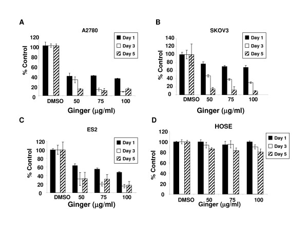
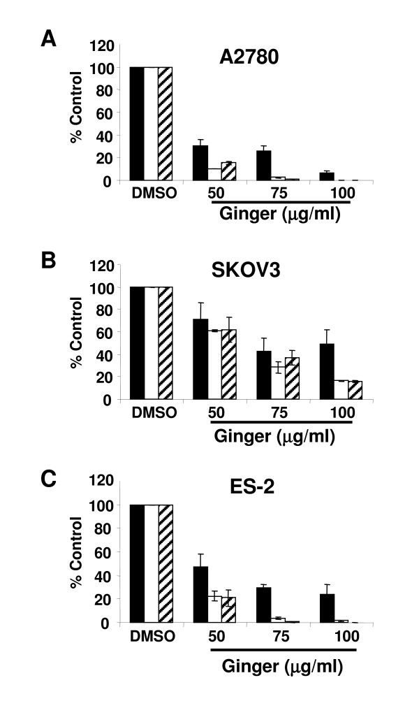
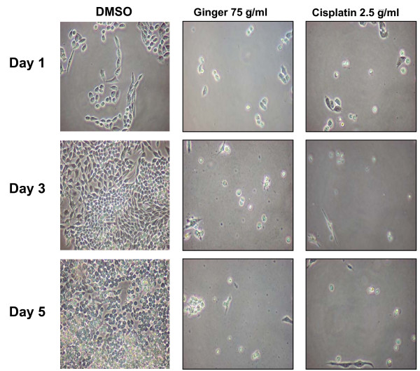
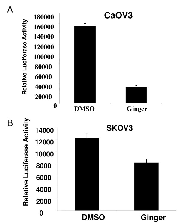
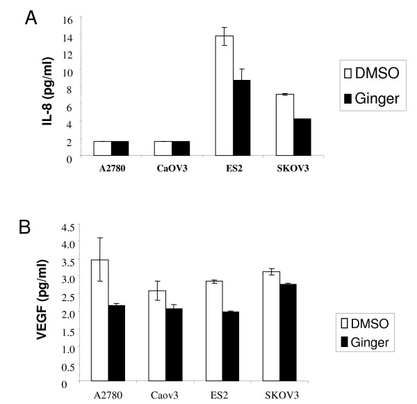
7.Ginger extract inhibits NFkB
8. Omega-3 Fatty Acids Inhibits NF-kB expression via PPARα-dependent pathway.
9. fish oil diet significantly increased the level of PTEN protein in the breast tumors.
In addition, the fish oil diet attenuated the PI 3 kinase and Akt kinase activity in the tumors leading to significant inhibition of NFκB activation. Fish oil diet also prevented the expression of anti-apoptotic proteins Bcl-2 and Bcl-XL in the breast tumors with concomitant increase in caspase 3 activity.
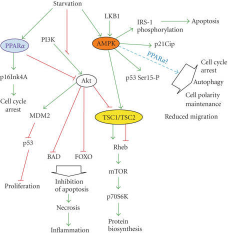
10.Docosahexaenoic Acid Induces Cell Death in Human Non-Small Cell Lung Cancer Cells by Repressing mTOR via AMPK Activation and PI3K/Akt Inhibition.
ERBB is abbreviated from erythroblastic oncogene B, a gene isolated from avian genome. It is also frequently called HER2 (from human epidermal growth factor receptor 2) or HER2/neu.
HER2 is a member of the human epidermal growth factor receptor (HER/EGFR/ERBB) family. Amplification or over-expression of this oncogenehas been shown to play an important role …
Wikipedia · Text under CC-BY-SA licenseNAC has been shown to be a potent inhibitor of NF-κB activation in vascular endothelial cells (Schubert et al. 2002). NAC and other antioxidants have been reported to inhibit hydrogen peroxide-induced NF-κB activation (Gupta et al. 2010). The central role played by NF-κB signal pathway in physiological and pathological conditions has made it a potential target for pharmacological intervention
Omega-3 polyunsaturated fatty acid promotes the inhibition of glycolytic enzymes and mTOR signaling by regulating the tumor suppressor LKB1
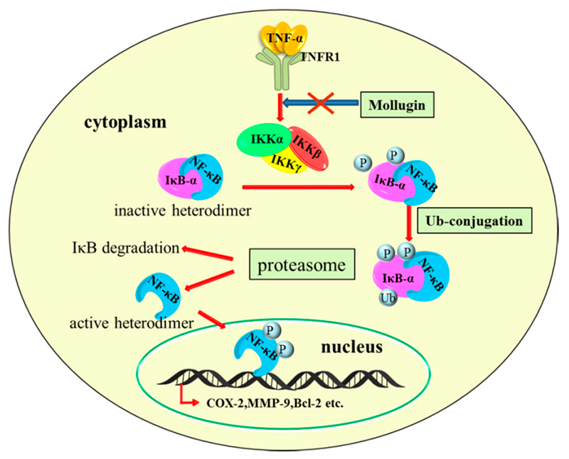
Figure 4. Inflammation and NF-κB signaling cross-talk is an important link in brain cancer pathogenesis. The inflammatory cytokines (TNF-α, IL-1β, IL-6, and IL-8) released in the tumor microenvironment generate signals that lead to activation of IκB kinases (IKKs). IKKs induce phosphorylation and subsequent proteosomal degradation of NF-κB inhibitor, IκBα(inhibitor of Kappa B). The resulting free NF-κB (e.g., heterodimer of p65 and p50 subunits) then translocates to the nucleus and activate the transcription of various target genes that encode anti-apoptotic proteins (e.g., B-cell lymphoma 2/ Bcl-2), pro-inflammatory cytokines and chemokines, adhesion molecules (integrins and cadherins), proteases (MMPs), DNA repair proteins such as the MGMT (O6-methylguanine DNA methyltransferase). Many of these regulators contribute significantly to the oncogenesis of gliomas.https://www.mdpi.com/2218-273X/7/2/34/htm
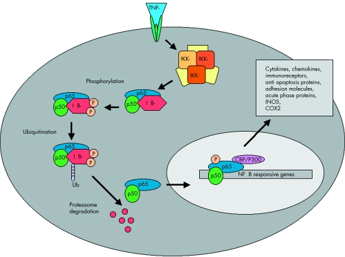
A model illustrating the signaling pathway of NFkB activation. NFkB, shown here as a heterodimer consisting of p65 and p50 subunits, regulates expression of a number of inflammatory genes. IKK is activated following brain ischemia, causing the phosphorylation of its inhibitor protein, IkB. IkB is phosphorylated by its corresponding kinase (IKK) leading to ubiquitination (ubi) and degradation in proteasomes. This causes the dissociation of NFkB from IkB. Liberated NFkB then translocates into the nucleus, turning on the transcription of its downstream target genes, IKK 5 IkB kinase
Docosahexaenoic Acid Induces Cell Death in Human Non-Small Cell Lung Cancer Cells by Repressing mTOR via AMPK Activation and PI3K/Akt Inhibition.
Affiliation: Department of Biochemistry, School of Medicine, Chungnam National University, Daejeon 301-747, Republic of Korea ; Infection Signaling Network Research Center, School of Medicine, Chungnam National University, Daejeon 301-747, Republic of Korea.
The anticancer properties and mechanism of action of omega-3 polyunsaturated fatty acids (ω3-PUFAs) have been demonstrated in several cancers; however, the mechanism in lung cancer remains unclear. Here, we show that docosahexaenoic acid (DHA), a ω3-PUFA, induced apoptosis and autophagy in non-small cell lung cancer (NSCLC) cells.
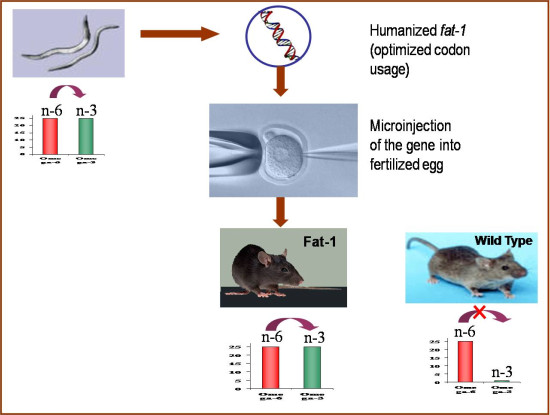
DHA-induced cell death was accompanied by AMP-activated protein kinase (AMPK) activation and inactivated phosphatidylinositol 3-kinase (PI3K)/Akt/mammalian target of rapamycin (mTOR) signaling. Knocking down AMPK and overexpressing Akt increased mTOR activity and attenuated DHA-induced cell death, suggesting that DHA induces cell death via AMPK- and Akt-regulated mTOR inactivation. This was confirmed in Fat-1 transgenic mice, which produce ω3-PUFAs. Lewis lung cancer (LLC) tumor cells implanted into Fat-1 mice showed slower growth, lower phospho-Akt levels, and higher levels of apoptosis and autophagy than cells implanted into wild-type mice. Taken together, these data suggest that DHA-induced apoptosis and autophagy in NSCLC cells are associated with AMPK activation and PI3K/Akt inhibition, which in turn lead to suppression of mTOR; thus ω3-PUFAs may be utilized as potential therapeutic agents for NSCLC treatment.
二十二碳六烯酸通过激活AMPK和抑制PI3K/Akt来抑制mTOR,从而诱导人非小细胞肺癌细胞死亡。
抗癌的属性和ω- 3多不饱和脂肪酸的作用机制(ω3-PUFAs)已经证明在一些癌症;然而,肺癌的机制尚不清楚。在这里,我们表明,二十二碳六烯酸(DHA),ω3-PUFA、诱导细胞凋亡和自噬在非小细胞肺癌(NSCLC)细胞。
dhad诱导的细胞死亡伴随着amp活化的蛋白激酶(AMPK)激活和失活的磷脂酰肌醇3-激酶(PI3K)/Akt/哺乳动物雷帕霉素靶蛋白(mTOR)信号通路。敲低AMPK和过表达Akt可增加mTOR活性,减弱DHA诱导的细胞死亡,提示DHA可通过AMPK和Akt调控的mTOR失活诱导细胞死亡。这是脂肪1证实转基因小鼠,产生ω3-PUFAs。与野生型小鼠相比,Lewis lung cancer (LLC)肿瘤细胞植入Fat-1小鼠后生长缓慢,磷酸化akt水平较低,凋亡和自噬水平较高。综上所述,这些数据表明dhad诱导的NSCLC细胞凋亡和自噬与AMPK激活和PI3K/Akt抑制有关,从而导致mTOR的抑制;因此ω3-PUFAs可能被利用作为一个潜在的治疗药物在非小细胞肺癌的治疗。
https://openi.nlm.nih.gov/detailedresult.php?img=PMC4538321_BMRI2015-239764.006&req=4
https://openi.nlm.nih.gov/detailedresult.php?img=PMC4538321_BMRI2015-239764.006&req=4https://openi.nlm.nih.gov/detailedresult.php?img=PMC4538321_BMRI2015-239764.006&req=4
NF-κB activation by tumour necrosis factor requires the Akt serine–threonine kinase | Nature
https://www.nature.com/articles/43466
Cardiolipin oxidation and cytochrome c release in the mitochondrial... | Download Scientific Diagram
https://www.researchgate.net/figure/Cardiolipin-oxidation-and-cytochrome-c-release-in-the-mitochondrial-pathway-to-apoptosis_fig1_275060022
过氧化物酶体增殖物激活受体α(PPAR-alpha),也称为NR1C1(核受体亚家族1,C组,成员1),是一种在人类中由PPARA基因编码的核受体蛋白。[5]与过氧化物酶体增殖物激活受体δ和过氧化物酶体增殖物激活受体γ一起,PPAR-α是过氧化物酶体增殖物激活受体亚家族的一部分。它是1990年斯蒂芬·格林(Stephen Green)克隆的PPAR家族的第一个成员,并已被确认为引起过氧化物酶体增殖的多种啮齿类肝癌致癌物的核受体。[6]
功能
PPAR-α是肝脏中脂质代谢的转录因子和主要调节剂。 PPAR-α在能量缺乏的条件下被激活,并且对于生酮过程是必需的,生酮过程是对长期禁食的关键适应性反应。[7] PPAR-α的激活通过上调与脂肪酸运输,脂肪酸结合和激活以及过氧化物酶体和线粒体脂肪酸β-氧化有关的基因来促进脂肪酸的吸收,利用和分解代谢。[8] PPAR-α主要通过配体结合而被激活。合成的配体包括用于治疗高血脂症的贝特类药物,以及各种不同的杀虫剂,除草剂,增塑剂和有机溶剂(统称为过氧化物酶体增殖剂)。内源性配体包括脂肪酸,例如花生四烯酸以及其他多不饱和脂肪酸,以及各种脂肪酸衍生的化合物,例如花生四烯酸代谢物的15-羟基二十二碳四烯酸家族的某些成员,例如。 15(S)-HETE,15(R)-HETE和15(S)-HpETE和13-羟基十八碳二烯酸,一种亚油酸代谢产物。组织分布
PPAR-α的表达在迅速氧化脂肪酸的组织中最高。在啮齿动物中,在肝脏和棕色脂肪组织中发现最高的PPAR-αmRNA表达水平,其次是心脏和肾脏。[9]在小肠和大肠,骨骼肌和肾上腺中发现较低的PPAR-α表达水平。人PPAR-α似乎在各种组织中更平等地表达,并在肝,肠,心脏和肾脏中高表达。
敲除研究
使用缺乏功能性PPAR-α的小鼠进行的研究表明,PPAR-α对于被称为过氧化物酶体增殖剂的各种合成化合物诱导过氧化物酶体增殖至关重要。[10]缺乏PPAR-α的小鼠对禁食的反应也受损,其特征是主要的代谢紊乱,包括血浆血浆中酮体水平低,低血糖和脂肪肝。[7]
药理
PPAR-α用作贝特类药物的细胞受体,贝特类药物是治疗血脂异常的一类药物。贝特类药物有效降低血清甘油三酸酯并提高血清HDL-胆固醇水平。[11]尽管已观察到贝特类药物治疗的临床益处,但与他汀类药物相比,总体结果好坏参半,导致人们对贝特类药物在冠状动脉心脏病的治疗中的广泛应用持保留态度。 PPAR-α激动剂可能具有治疗非酒精性脂肪肝的治疗价值。 PPAR-α也可能是某些抗惊厥药的作用部位。[12] [13]
靶基因
PPAR-α通过改变大量靶基因的表达来控制生物学过程。因此,PPAR-α的功能作用与其靶基因的生物学功能直接相关。基因表达谱研究表明,PPAR-alpha靶基因有数百种。[8] PPAR-alpha的经典靶基因包括PDK4,ACOX1和CPT1。低通量和高通量基因表达分析已经创建了完整的图谱,该图谱说明了PPAR-α通过调节涉及脂质代谢各个方面的众多基因而作为脂质代谢的主要调节剂的作用。这些针对小鼠肝脏和人类肝脏构建的地图将PPAR-α置于影响脂肪酸摄取和细胞内结合,线粒体β-氧化和过氧化物酶体脂肪酸氧化,生酮作用,甘油三酸酯更新,糖异生和胆汁合成的调节中心/分泌。
Omega-3脂肪酸(FAs)是过氧化物酶体增殖物激活受体-α(PPARα)的天然配体,可以激活PPARα
Omega-3 fatty acids (FAs) are natural ligands of the peroxisome proliferator-activated receptor-α (PPARα)
Omega-3脂肪酸(FAs)是过氧化物酶体增殖物激活受体-α(PPARα)的天然配体(Omega-3 fatty acids (FAs) are natural ligands of the peroxisome proliferator-activated receptor-α (PPARα)),后者是一种核受体,可调节参与脂质代谢的基因的表达水平。 PPARα基因的L162V多态性与代谢状况恶化有关。我们假定与L162纯合子相比,与O162-3 FA孵育后,携带PPARα-V162等位基因的受试者在PPARα及其靶基因的表达上存在差异。将六名携带年龄和体重指数配对的PPARα-V162等位基因与六个L162纯合子配对的六名男性的外周血单核细胞分化为巨噬细胞,并用二十碳五烯酸(EPA),二十二碳六烯酸(DHA)或EPA:DHA的混合物活化。数据表明,与添加DHA和EPA:DHA混合物后的L162纯合子相比,PPARα-V162等位基因携带者的PPARα和载脂蛋白AI(APOA1)的基因表达水平显着降低。此外,添加EPA:DHA混合物后,脂蛋白脂肪酶(LPL)基因表达在PPARαL162V多态性亚组中表现出较低的趋势。因此,携带PPARα-V162等位基因的个体可能因响应omega-3 FA补充而引起的基因表达率的改变而显示出脂质水平的改善。Omega-3脂肪酸(FAs),尤其是二十碳五烯酸(EPA)和二十二碳六烯酸(DHA),对心血管有好处。Omega-3脂肪酸对脂质代谢的影响可能是由基因表达的改变介导的。更具体地说,Omega-3脂肪酸及其衍生物是(PPARα的天然配体,二聚化的类维生素a×受体(R×R)之前触发目标基因的表达[13]。特定的靶基因包括脂蛋白脂肪酶(LPL)[15](甘油三酸酯代谢的中心酶)和载脂蛋白AI (APOAI)[34](高密度脂蛋白的关键结构元素)。总的来说,欧米伽- 3 FAs绑定到PPARα因此有可能降低血浆甘油三酯(TG)水平和增加脂蛋白胆固醇(高密度脂蛋白胆固醇)的水平。
Omega-3 fatty acids regulate gene expression levels differently in subjects carrying the PPARα L162V polymorphism
https://www.ncbi.nlm.nih.gov/pmc/articles/PMC2745745/
图3:PPARα通过抑制主要的炎性转录因子NFκB和AP-1拮抗主要的炎性信号通路。此外,PPARα通过上调解偶联蛋白UCP2和UCP3减少ROS介导的炎症。有关更多详细说明,请参见文本。箭头表示激活/上调,而虚线表示细胞蛋白或过程的抑制/下调。 Erk1 / 2-细胞外信号应答激酶1/2; IκB-NFκB抑制剂; MAPK-促分裂原活化蛋白激酶; NFκB—核因子κB; ROS-活性氧。
NFKB mechanism of action - NF-κB - Wikipedia
https://en.wikipedia.org/wiki/NF-%CE%BAB#/media/File:NFKB_mechanism_of_action.pngNF-κB(活化的B细胞的核因子κ轻链增强子)是一种蛋白质复合物,可控制DNA的转录,细胞因子的产生和细胞存活。 NF-κB几乎存在于所有动物细胞类型中,并参与细胞对刺激的反应,例如应激,细胞因子,自由基,重金属,紫外线照射,氧化的LDL以及细菌或病毒抗原。NF-κB在调节对感染的免疫反应中起关键作用。 NF-κB调节适当与癌症,炎性和自身免疫性疾病,败血症性休克,病毒感染和免疫发育不当有关。 NF-κB也与突触可塑性和记忆过程有关。
NF-κB作用机理 在该图中,以Rel和p50蛋白组成的NF-κB异二聚体为例。处于失活状态时,NF-κB位于与抑制蛋白IκBα复合的胞质溶胶中。通过整合膜受体的介导,多种细胞外信号可以激活酶IκB激酶(IKK)。 IKK反过来会磷酸化IκBα蛋白,从而导致泛素化,IκBα与NF-κB的分离以及蛋白酶体最终降解IκBα。然后,活化的NF-κB易位到细胞核中,并与称为响应元件(RE)的特定DNA序列结合。然后,DNA /NF-κB复合物募集其他蛋白质,例如共激活因子和RNA聚合酶,它们将下游DNA转录成mRNA。反过来,mRNA被翻译成蛋白质,导致细胞功能改变。
维生素C调节信号响应的示意图。维生素C作为DHA通过葡萄糖转运蛋白进入细胞,并迅速还原为AA。 ROS通过激活IKKβ诱导NF-κB信号传导应答,而AA猝灭ROS,抑制IKKβ的激活。在这些过程中,AA氧化成DHA,而DHA抑制IKKβ。
氧化的Omega-3脂肪酸通过PPARα依赖性途径抑制NF-κB活化
Oxidized Omega-3 Fatty Acids Inhibit NF-κB Activation Via a PPARα-Dependent Pathway
动脉硬化、血栓形成与血管生物学。2004;24:1621-1627
摘要目的—
这项研究的目的是确定氧化的和天然的omega-3脂肪酸对趋化因子MCP-1和IL-8内皮表达的影响,并且,如果有效抑制趋化因子表达,则确定抑制趋化因子表达的机制。
方法和结果-
使用酶联免疫吸附测定法,我们表明氧化的EPA和DHA而非未氧化的EPA或DHA抑制细胞因子诱导的单核细胞趋化蛋白(MCP)-1的内皮表达,并在较小程度上抑制IL-8。在电泳迁移率变动分析中,氧化的EPA而非未氧化的EPA可以有效抑制细胞因子诱导的内皮细胞核因子-κB(NF-κB)的活化。使用蛋白质印迹分析,我们表明抑制NF-κB活化不是由预防IκBα的磷酸化引起的,因为氧化的EPA不能抑制细胞因子诱导的IκBα的磷酸化和泛素化。此外,氧化的EPA抑制了野生型小鼠内皮细胞中NF-κB的激活,但对过氧化物酶体增殖物激活受体α(PPARα)缺陷的小鼠中内皮细胞中NF-κB的激活没有抑制作用,表明氧化的EPA需要PPARα来抑制NF-κB。
结论—
这些研究表明,鱼油的抗炎作用可能是由于氧化的omega-3脂肪酸通过PPARα依赖性途径对NF-κB活化的抑制作用所致。
讨论
在我们以前的研究中,我们注意到氧化的EPA是PPARα的有效激活剂,而氧化的EPA对白细胞与内皮的相互作用的抑制作用需要PPARα,12因此我们推测氧化的EPA可能抑制细胞因子诱导的NF-κB。通过PPARα依赖性途径激活。在这里,我们表明,虽然氧化的EPA抑制了野生型细胞中细胞因子诱导的NF-κB的活化,但对PPARα缺乏的内皮细胞没有抑制作用,这表明氧化的EPA通过PPARα介导了对NF-κB的抑制作用。 PPARα介导的氧化EPA对NF-κB活化的抑制作用可能是PPARα与p50 / p65亚基的直接相互作用。 Delerive等人(1999年)使用谷胱甘肽S-转移酶下拉实验表明,在纤维蛋白(PPARα激动剂)处理后,PPARα在体外与p65发生物理相互作用[33]。
与我们之前的研究[11,12]一起,我们发现omega-3脂肪酸的自氧化作用导致生成具有强抗炎特性的氧化化合物,这些化合物可抑制促炎反应,如白细胞粘附受体和趋化因子的表达。氧化的omega-3脂肪酸的抗炎作用的中心主题可能是通过PPARα依赖性途径抑制NF-κB。 ω-3脂肪酸的氧化很可能发生在由氧化酶(例如NADPH氧化酶,髓过氧化物酶,环加氧酶,脂氧合酶)表达增加引起的活动性炎症区域,并在这些区域产生活性氧氧化性PUFA。这些产品的鉴定可产生具有有效PPARα激动剂和抗NF-κB特性的有效,低毒,促炎反应抑制剂,用于治疗炎性疾病。
Oxidized Omega-3 Fatty Acids Inhibit NF-κB Activation Via a PPARα-Dependent Pathway
Archana Mishra , Ashok Chaudhary , and Sanjeev Sethi Originally published1 Jul 2004https://doi.org/10.1161/01.ATV.0000137191.02577.86
Abstract
Objective— The aim of this study was to determine the effects of oxidized versus native omega-3 fatty acids on the endothelial expression of chemokines MCP-1 and IL-8, and, if effective in inhibiting chemokine expression, to determine the mechanism for the inhibition of chemokine expression.
Methods and Results— Using enzyme-linked immunosorbent assays, we show that oxidized EPA and DHA but not unoxidized EPA or DHA inhibit cytokine-induced endothelial expression of monocyte chemoattractant protein (MCP)-1 and, to a lesser extent, IL-8. In electrophoretic mobility shift assays, oxidized EPA but not unoxidized EPA potently inhibited cytokine-induced activation of endothelial nuclear factor-κB (NF-κB). Using Western blot analyses, we show that the inhibition of NF-κB activation was not caused by prevention of phosphorylation of IκBα because oxidized EPA did not inhibit cytokine-induced phosphorylation and ubiquination of IκBα. Furthermore, oxidized EPA inhibited NF-κB activation in endothelial cells derived from wild-type mice but had no inhibitory effects on NF-κB activation in endothelial cells derived from peroxisome proliferator-activated receptor α (PPARα)-deficient mice, indicating that oxidized EPA requires PPARα for its inhibitory effects on NF-κB.
Conclusions— These studies show that the antiinflammatory effects of fish oil may result from the inhibitory effects of oxidized omega-3 fatty acids on NF-κB activation via a PPARα-dependent pathway.Discussion
Omega-3 fatty acids in fish oil has been reported to improve the prognosis of several chronic inflammatory diseases characterized by leukocyte accumulation, including atherosclerosis, inflammatory bowel disease, rheumatoid arthritis, psoriasis, etc.1–4
Omega-3 fatty acids, such as EPA and DHA, are highly polyunsaturated and readily undergo oxidation at ambient and subambient temperatures, even in the absence of exogenous oxidizing reagents.5,6 In view of the ease with which omega-3 PUFA spontaneously oxidize and in vivo data suggesting extensive accumulation of oxidation products after fish oil consumption, we investigated the possibility that oxidized omega-3 fatty acids may be an important component of the observed antiinflammatory effects of fish oil. In our previous studies, we showed that oxidized EPA and not unoxidized EPA pretreatment of HUVEC inhibits leukocyte adhesion to cytokine-stimulated HUVEC, and this effect is mediated through a PPARα-dependent pathway.11,12 In our present studies, we extend these observations and show that oxidized EPA is also effective in inhibiting cytokine-induced endothelial chemokine expression, particularly MCP-1, and propose a mechanism for these antiinflammatory effects.
The fact that oxidation of the omega-3 fatty acids is required for the aforementioned antiinflammatory effects is also pointed out by other studies. Nohe et al and De Caterina et al have shown that prolonged incubation of endothelial cells with native omega-3 fatty acids (26 hours of total exposure, 6 hours before and 20 hours after TNFα stimulation) results in inhibition of cytokine and adhesion molecule expression, whereas shorter incubation periods (6 hours) has no effect.30–32 This suggests that oxidation of omega-3 fatty acids takes place during the long incubation period, and that the oxidation product(s) and not the native omega-3 fatty acids are most likely responsible for the inhibition of leukocyte–endothelial interactions.
To ascertain that the inhibition of chemokine expression was not caused by the cytotoxic effects of oxidized EPA, we performed MTT assays, which showed that oxidized EPA in the doses used for our experiments had no effect on the viability of endothelial cells (data not shown). Also, oxidized EPA did not have any effect on the constitutively expressed surface proteins such as von Willebrand factor, endoglin, and human leukocyte antigen class I molecules.11,12
The expression of MCP-1 and IL-8 is regulated through activation of NF-κB. NF-κB activation appears to be necessary for the induction of chemokine genes, and deletion of NF-κB binding sites results in an inability to induce these genes.26 In these studies, we show that oxidized EPA and not unoxidized EPA inhibits cytokine-induced activation of NF-κB and promotes cytosolic retention of the p50 and p65 subunits. Hence, the oxidized EPA-mediated inhibition of MCP-1 and IL-8 in cytokine-stimulated endothelial cells could be explained by an inhibitory effect of oxidized EPA on NF-κB activity.
The difference in the extent of inhibition of MCP-1 and IL-8 expression by oxidized EPA is most likely caused by the differential effects of oxidized EPA on endothelial NF-κB and AP-1 activation. Oxidized EPA almost completely inhibits endothelial NF-κB activation, whereas it had minimal effects in preventing AP-1 activation. Because AP-1 activation alone can result in IL-8 expression,28 oxidized EPA had only a mild inhibitory effect on IL-8 expression.
What is the mechanism for the oxidized EPA-mediated inhibition of cytokine-induced NF-κB activation? We first hypothesized that oxidized EPA inhibits phosphorylation of IκBα by inhibiting the IKK (kinase) complex. However, it is unlikely that oxidized EPA inhibits IKK (kinase) activity because phosphorylation and ubiquination of IkBα is noted in oxidized EPA-treated endothelial cells. In fact, when normalized to actin controls, oxidized EPA pretreatment before TNFα stimulation for 60 minutes resulted in 30% more p-IkBα when compared with TNFα-treated cells. Also, it unlikely that oxidized EPA induces IkBα expression because after 15 minutes of cytokine stimulation, no IkBα is noted in the cytoplasm of cells pretreated with oxidized EPA. After 60 minutes, IκBα starts to appear in the cytoplasm of oxidized EPA pretreated cells and its concentration is, in fact, somewhat less than the TNFα-treated cells. Thus, it is likely that oxidized EPA does not prevent NF-κB activation by increasing the expression of IκBα.DISCUSSION
In our previous studies we noted that oxidized EPA is a potent activator of PPARα and that PPARα is needed for the inhibitory effects of oxidized EPA on leukocyte–endothelial interactions,12 which lead us to hypothesize that oxidized EPA might inhibit cytokine-induced NF-κB activation through a PPARα-dependent pathway. Here, we show that although oxidized EPA inhibits cytokine-induced NF-κB activation in wild-type cells, it has no inhibitory effects in PPARα-deficient endothelial cells, suggesting that oxidized EPA mediates its inhibitory effects on NF-κB through PPARα. The PPARα-mediated inhibitory effects of oxidized EPA on NF-κB activation are possibly through direct interactions of PPARα with the p50/p65 subunits. Delerive et al (1999) have shown, using glutathione S-transferase pull-down experiments, that after fibrate (PPARα agonist) treatment, PPARα physically interacts with p65 in vitro.33
Taken together with our previous studies,11,12 we show that auto-oxidation of omega-3 fatty acids results in the generation of oxidized compounds with potent antiinflammatory properties that inhibit proinflammatory responses such as leukocyte adhesion receptor and chemokine expression. The central theme for the antiinflammatory effects of oxidized omega-3 fatty acids is likely through inhibition of NF-κB via a PPARα-dependent pathway. The oxidation of omega-3 fatty acids is likely to occur in areas of active inflammation caused by increased expression of oxidative enzymes (eg, NADPH oxidase, myeloperoxidase, cyclooxygenase, lipoxygenase) and the generation of reactive oxygen species in these areas, which are capable of oxidizing PUFAs. The identification of these products could result in potent, low-toxicity, proinflammatory response inhibitors with potent PPARα agonist and anti-NF-κB properties for the treatment of inflammatory diseases.
Oxidized Omega-3 Fatty Acids Inhibit NF-κB Activation Via a PPARα-Dependent Pathway | Arteriosclerosis, Thrombosis, and Vascular Biology
https://www.ahajournals.org/doi/10.1161/01.atv.0000137191.02577.86
口服补充Omega3脂肪酸可抑制慢性淋巴细胞白血病(CLL)细胞中的NFkB活化
Oral Supplementation with Omega3 Fatty Acids Inhibits NFkB Activation In Chronic Lymphocytic Leukemia (CLL) Cells.
Oscar F. F Ballester
简介:核因子κB(NFkB)是参与CLL细胞生长和存活的关键转录因子。 NFkB被认为是开发用于治疗各种恶性肿瘤的新疗法的重要目标。在体外和实验动物模型中,已证明补充OMEGA-3脂肪酸(O3FA)可抑制NFkB活性。患者和方法:患有早期CLL(Rai 0-II期)且无需治疗的患者,该患者已纳入I-II期试验。 O3FA补充剂总共服用12个月,剂量范围为2250 mg(EPA加DHA),每日可耐受的剂量增加到4500 mg和6750 mg。在通过梯度离心分离单个核细胞后,在外周血样品中测量NFkB活性,并以发光单位/μg蛋白质表示。在研究期间获得基线和多个系列样品。使用标准的LD50方法对阿霉素进行体外细胞毒性试验。通过气相色谱分析红细胞和淋巴细胞膜脂质组成来监测依从性。
结果:15名患者被纳入该试验,其中8名目前已经完成了计划的研究期12个月。没有发现疾病活动的显着临床变化。 O3FA的耐受性良好。补充导致红细胞和淋巴细胞膜的O3FA成分呈剂量依赖性增加。在基线时,CLL患者的NFkB高于正常对照者观察到的范围(2.05×10⁴至2.32×10⁵ NFkB流明单位/微克)。基线时CLL患者的中位数为11.60×106
Oral Supplementation with Omega3 Fatty Acids Inhibits NFkB Activation In Chronic Lymphocytic Leukemia (CLL) Cells.
Oscar F. F Ballester
Introduction: Nuclear factor kappa B (NFkB) is a critical transcription factor involved in the growth and survival of CLL cells. NFkB is recognized as an important target for the development of novel therapies for the treatment of various malignancies. In vitro and in experimental animal models, OMEGA-3 fatty acid (O3FA) supplementation has been shown to inhibit NFkB activity. Patients and Methods: Patients with early stage CLL (Rai stages 0-II) who required no therapy, where accrued to this phase I-II trial. O3FA supplements were given for a total of 12 months at doses ranging from 2250 mg (EPA plus DHA), escalated to 4500 mg and 6750 mg per day as tolerated. NFkB activity was measured in peripheral blood samples after separation of mononuclear cell by gradient centrifugation and expressed as luminescence units/μ g of protein. Baseline and multiple serial samples were obtained during the study period. In-vitro cytotoxicity assays to doxorubicin were conducted using standard LD50 methods. Compliance was monitored by analysis of red cell and lymphocyte membrane lipid composition by gas chromatography. Results: Fifteen patients have been accrued to the trial, 8 of them have currently completed the planned 12 months of the study period. No significant clinical changes in disease activity were noted. O3FA was well tolerated. Supplementation resulted in a dose-dependent increase of O3FA composition of red cell and lymphocyte membranes in a dose dependent manner. At baseline, CLL patients had NFkB above the range observed in normal controls (2.05 × 10⁴ to 2.32 × 10⁵ NFkB lum units/μ g). The median value in CLL patients at baseline was 11.60 × 10⁶
Oral Supplementation with Omega3 Fatty Acids Inhibits NFkB Activation In Chronic Lymphocytic Leukemia (CLL) Cells. | Blood | American Society of Hematology
https://ashpublications.org/blood/article/116/21/4607/66865/Oral-Supplementation-with-Omega3-Fatty-Acids
NF-κB inhibitors and activators
Use this guide to find the right compound to activate or inhibit the NF-κB signaling pathway.
Multiple pharmacological compounds can activate or inhibit proteins involved in NF-kB signaling regulation. The table below highlights some of the small molecules that can be used to study NF-kB pathway.
Antioxidants
Antioxidants such as PDTC40 and NAC41 have shown potential to inhibit NF-κB activation either by exogeneous induction (e.g. LPS, TNFα) or hydrogen peroxide treatment. Antioxidants are likely inhibit NF-κB by scavenging reactive oxygen intermediates involved in the NF-κB pathway42.
Anti-inflammatory and immunosuppressant drugs
The commonly available NSAID, sodium salicylate was shown to bind IKKβ43 and inhibit proteasome activity44 potentially reducing IκB degradation. A potent immunomodulatory glucocorticoid steroid, dexamethasone (DEX) exhibited interference with NF-κB activation and reduced TNFα production45,46.
Investigations into immunosuppressant drugs revealed cyclosporin A (CsA) to inhibit NF-κB /RelA activation and block IL-2 and IL-8 gene expression47,48. FK506 (tacrolimus) another commercially available immunosuppressive drug blocks p50 nuclear translocation thereby reducing activation of its subsequent promoters and gene expression of inflammatory cytokines such as IL-249.
NF-kB inhibitors and activators | Abcam
https://www.abcam.com/reagents/nf-kb-small-molecule-guide
Two faces of PPARα/NFκB signaling pathway in inflammatory responses to adipocytes lipolysis in grass carp Ctenopharyngodon idella
College of Animal Science and Technology, Northwest A&F University, Yangling, 712100, China
Highlights
•
Lipolysis has a dose-dependent differential impact on inflammatory response in adipocytes.
•
PPARα inhibited NF-κB signaling pathway in grass carp adipocytes.
•
Excess lipolysis produces ROS that contributes to adipocyte inflammation.
Abstract
Adipose tissue plays an important role in energy reservation, also be considered as vital immunological organ in animals. Adipocytes are the basic unit of adipose tissue, while little is known about the relationship between lipid metabolism and inflammatory response in fish adipocytes so far. In this study, forskolin was used to induce adipocyte lipolysis, and 5 μM forskolin and 30 μM forskolin both triggered lipolysis by increasing ATGL expression. Consequently, 30 μM Forskolin instead of 5 μM Forskolin induced the expression of NF-κB and its target pro-inflammatory cytokine genes including MCP-1, IL-6 and TNF-α. Further study found that low grade rate of lipolysis activated PPARα gene, and its inhibitory effect on the mRNA expression of NF-κB and its target genes inhibited the adipocyte inflammation. On the contrary, high grade rate of lipolysis increased the expression levels of NF-κB and its target genes, while their expression were attenuated by inhibition of reactive oxygen species (ROS) using α-tocopherol, suggesting that ROS generated due to the PPARα-mediated oxidation of released fatty acids from lipolysis may contribute to adipocyte inflammation. These results indicated that PPARα has dose effect in inflammatory responses to adipocyte lipolysis in grass carp. Taken together, grass carp adipocytes have immune activity. The inflammatory response is linked to the grade rate of adipocyte lipolysis in grass carp adipocytes, and excessive adipocyte lipolysis may promote a dynamic immune response in adipose tissue. This is the first study showing the regulatory effects of lipolysis on immune functions in fish adipocytes.
Two faces of PPARα/NFκB signaling pathway in inflammatory responses to adipocytes lipolysis in grass carp Ctenopharyngodon idella - ScienceDirect
https://www.sciencedirect.com/science/article/abs/pii/S1050464819303006
NF-κB信号传导在慢性炎性气道疾病中的作用
NF-kappaB Signaling in Chronic Inflammatory Airway Disease
通过迈克尔·舒利加(Michael Schuliga)
墨尔本大学肺部健康研究中心(LHRC),墨尔本大学Grattan St.,Parkville 3010,澳大利亚维多利亚
抽象
哮喘和慢性阻塞性肺疾病(COPD)是阻塞性气道疾病,其根本病因和表型不同,但药理治疗方式重叠。在哮喘和COPD中,氧化应激通过诱导炎症基因表达而导致气道炎症。氧化还原敏感的转录因子,核因子(NF)-κB(NF-κB),是调节呼吸道病理中细胞因子活性的各种炎症网络的重要参与者。糖皮质激素(GCs)(哮喘的主要治疗药物)的抗炎作用涉及抑制NF-κB诱导的基因转录。配体结合的GC受体(GRs)与NF-κB结合以抑制NF-κB响应基因的转录(即反转录)。然而,在重度哮喘和COPD中,由于翻译后GR和染色质重塑组蛋白的变化,GC导致的NF-κB的反式表达被否定。靶向NF-κB活化的疗法,包括IκB激酶(IKKs)抑制剂,可能是哮喘和COPD的潜在疗法。此外,逆转GR /组蛋白乙酰化显示出有望通过增强NF-κB抑制来治疗类固醇难治性气道疾病。这篇综述检查了气道炎症中的NF-κB信号传导及其作为治疗哮喘和COPD靶标的潜力。
NF-kappaB Signaling in Chronic Inflammatory Airway Disease
by Michael Schuliga
Lung Health Research Centre (LHRC), Department Pharmacology and Therapeutics, University of Melbourne, Grattan St., Parkville 3010, Victoria, Australia
Abstract
Asthma and chronic obstructive pulmonary disease (COPD) are obstructive airway disorders which differ in their underlying causes and phenotypes but overlap in patterns of pharmacological treatments. In both asthma and COPD, oxidative stress contributes to airway inflammation by inducing inflammatory gene expression. The redox-sensitive transcription factor, nuclear factor (NF)-kappaB (NF-κB), is an important participant in a broad spectrum of inflammatory networks that regulate cytokine activity in airway pathology. The anti-inflammatory actions of glucocorticoids (GCs), a mainstay treatment for asthma, involve inhibition of NF-κB induced gene transcription. Ligand bound GC receptors (GRs) bind NF-κB to suppress the transcription of NF-κB responsive genes (i.e., transrepression). However, in severe asthma and COPD, the transrepression of NF-κB by GCs is negated as a consequence of post-translational changes to GR and histones involved in chromatin remodeling. Therapeutics which target NF-κB activation, including inhibitors of IκB kinases (IKKs) are potential treatments for asthma and COPD. Furthermore, reversing GR/histone acetylation shows promise as a strategy to treat steroid refractory airway disease by augmenting NF-κB transrepression. This review examines NF-κB signaling in airway inflammation and its potential as target for treatment of asthma and COPD.
Keywords: airway smooth muscle; allergic asthma; alveolar macrophages; cigarette smoke; eosinophils; epithelium; emphysema; histone deacetylase; phosphoinositide 3 kinase-delta (PI3K-δ); sirtuins
Biomolecules | Free Full-Text | NF-kappaB Signaling in Chronic Inflammatory Airway Disease
https://www.mdpi.com/2218-273X/5/3/1266
维生素C是一种激酶抑制剂:脱氢抗坏血酸抑制IκBα激酶β,抑制NF-kB的激活
Vitamin C Is a Kinase Inhibitor: Dehydroascorbic Acid Inhibits IκBα Kinase β
Department of Clinical Laboratories,1 Program in Molecular Pharmacology and Chemistry, Memorial Sloan-Kettering Cancer Center,2 Structural Biology Program, Department of Physiology and Biophysics, Mount Sinai School of Medicine, New York, New York3
*Corresponding author. Mailing address: Program in Molecular Pharmacology and Chemistry, Box 451, Memorial Sloan-Kettering Cancer Center, New York, NY 10021. Phone: (212) 639-8483. Fax: (212) 772-8589. E-mail: gro.ccksm.iks@edlog-d.
ABSTRACT
Reactive oxygen species (ROS) are key intermediates in cellular signal transduction pathways whose function may be counterbalanced by antioxidants. Acting as an antioxidant, ascorbic acid (AA) donates two electrons and becomes oxidized to dehydroascorbic acid (DHA). We discovered that DHA directly inhibits IκBα kinase β (IKKβ) and IKKα enzymatic activity in vitro, whereas AA did not have this effect. When cells were loaded with AA and induced to generate DHA by oxidative stress in cells expressing a constitutive active IKKβ, NF-κB activation was inhibited. Our results identify a dual molecular action of vitamin C in signal transduction and provide a direct linkage between the redox state of vitamin C and NF-κB signaling events. AA quenches ROS intermediates involved in the activation of NF-κB and is oxidized to DHA, which directly inhibits IKKβ and IKKα enzymatic activity. These findings define a function for vitamin C in signal transduction other than as an antioxidant and mechanistically illuminate how vitamin C down-modulates NF-κB signaling.维生素C是一种激酶抑制剂:脱氢抗坏血酸抑制IκBα激酶β
摘要
活性氧(ROS)是细胞信号转导通路中的关键中间体,其功能可能会被抗氧化剂抵消。抗坏血酸(AA)充当抗氧化剂,提供两个电子并被氧化成脱氢抗坏血酸(DHA)。我们发现DHA在体外直接抑制IκBα激酶β(IKKβ)和IKKα的酶活性,而AA则没有这种作用。当在细胞中加载AA并通过表达本构活性IKKβ的细胞中的氧化应激诱导生成DHA时,NF-κB激活受到抑制。我们的结果确定了维生素C在信号转导中的双重分子作用,并揭示了维生素C的氧化还原状态与NF-κB信号事件之间的直接联系。 AA淬灭参与NF-κB活化的ROS中间体,并被氧化成DHA,直接抑制IKKβ和IKKα的酶活性。这些发现定义了维生素C在信号传导中的功能,除了作为抗氧化剂外,还从机理上阐明了维生素C如何下调NF-κB信号传导。
饮食中的维生素C对人类,灵长类动物,豚鼠和其他几种缺少l-古洛诺-γ-内酯氧化酶(它们是葡萄糖从生物合成途径中的最终酶)的动物和昆虫至关重要(25)。在生理条件下,维生素C主要以还原形式存在,即抗坏血酸(AA);它还以氧化形式脱氢抗坏血酸(DHA)微量存在。有两种已知的运输维生素C的机制(21)。存在于所有细胞中的通用系统通过便利的葡萄糖转运蛋白将维生素C作为DHA转运(34)。进入细胞后,DHA会迅速还原并以AA的形式累积(34、35)。第二种转运系统在专门的细胞中起作用,其中AA通过钠依赖性AA共转运蛋白(SVCT1和/或SVCT2)直接转运到细胞中(33)。
AA是胶原蛋白(26),肉碱(28)和去甲肾上腺素(18)以及激素酰胺化(8)的生物合成酶的辅助因子。在血浆和细胞中,AA是一种强大的抗氧化剂,可淬灭活性氧(ROS)和活性氮(10,14)。细胞内维生素C可以预防细胞死亡并抑制氧化应激诱导的突变(12,22,37)。在淬灭自由基的过程中,抗坏血酸会提供一个电子,成为不稳定的中间体抗坏血酸自由基,可逆地还原为抗坏血酸。抗坏血酸基团可以提供第二个电子并转化为DHA(13,14)。 DHA可能还原为AA或不可逆地水解为2,3-二酮-古洛糖酸,然后被代谢为苏糖酸和草酸(14)。在装有AA并暴露于过氧化氢的细胞中,AA转化为DHA,其中一些通过葡萄糖转运蛋白从细胞中流出,从而提供了一种将维生素C循环到细胞外培养基中的机制(12)。或者,可将细胞内DHA转运至细胞内区室和细胞器(2,20)。 DHA主要起维生素C的易运输形式的作用(36)。ROS作为化学第二信使分子在细胞反应中起关键作用,反之,抗氧化剂调节选定的信号反应(24)。例如,ROS激活转录因子,例如NF-κB,在宿主防御,炎症和凋亡中很重要(1、11、32)。促炎细胞因子,例如肿瘤坏死因子α(TNF-α),过氧化氢和神经酰胺,通过诱导IκB蛋白的磷酸化来激活NF-κB(11,19)。磷酸化的IκBα释放NF-κB,并通过蛋白酶体途径自身降解(17),而未磷酸化的IκB与胞浆中的NF-κB结合,阻止其核迁移。最初有报道说,AA通过激活p38丝裂原活化的蛋白激酶(MAPK)抑制内皮细胞中TNF-α诱导的NF-κB的活化(4)。但是,我们最近发现AA通过抑制与IκBα磷酸化有关的激酶的激活来抑制TNF-α依赖性的NF-κB激活(6)。
我们研究了维生素C对NF-κB激活的调节,发现DHA在体外和细胞试验中直接抑制了IKKβ和IKKα的激酶活性。因此,我们的数据表明维生素C调节NF-κB功能的双重作用机制。首先,作为抗氧化剂猝灭ROS,AA抑制ROS介导的信号转导事件。其次,在氧化成DHA后,维生素C直接抑制IKK激酶活性。材料和方法
维生素C负载。
如前所述(6),HeLa细胞中装有维生素C。简而言之,将细胞与孵育缓冲液(15 mM HEPES [pH 7.4],135 mM NaCl,5 mM KCl,1.8 mM CaCl2、0.8 mM MgCl2)(pH 7.4)孵育30分钟,然后用不同浓度的DHA处理30分钟在相同缓冲液中于37°C放置3分钟。 DHA获自Sigma(美国密苏里州圣路易斯)或通过将AA与抗坏血酸氧化酶(Sigma)孵育酶促产生。
免疫印迹分析。
如前所述制备细胞提取物(6)。使用以下兔多克隆抗体进行了免疫印迹分析:抗磷酸化IκBα,抗IκBα,抗p38 MAPK,抗磷酸化p38 MAPK,抗磷酸化的p44 / 42 MAPK(Cell Signaling Technology,Beverly,MA)。抗p44 / 42 MAPKs(纽约州普莱西德湖的Upstate Biotech)和抗FLAG抗体(Sigma)。将膜与辣根过氧化物酶偶联的抗兔免疫球蛋白G抗体一起温育,并使用增强的化学发光测定法(Amersham Pharmacia Biotech,Piscataway,N.J。)显示蛋白质。
转染和荧光素酶测定。
用pNFκB-luc(Clontech,Palo Alto,CA)瞬时转染HeLa细胞或将其与含IKKβ(SS / EE)(组成性活性IKKβ的丝氨酸177和181分别被谷氨酸替代)的质粒共转染通过磷酸钙法或Superfect(QIAGEN,瓦伦西亚,加利福尼亚)。将细胞与每毫升30 ngTNF-α一式三份孵育,时间为图中所示。使用萤光素酶报告基因测定系统(Promega)测定萤光素酶活性。激酶测定。
通过用抗IKKβ抗体(Santa Cruz Biotechnology,Santa Cruz,CA)进行免疫沉淀从TNF-α处理的细胞提取物中分离出内源性IKKβ。从抗FLAG抗体珠(Sigma)的转染HeLa细胞提取物中分离出IKKβ(SS / EE)及其突变体。从转染的293T细胞中分离出重组IKKα和IKKβ。用Genejammer转染试剂(Stratagene,La Jolla,CA)用含有IKKα或IKKβ的质粒瞬时转染细胞。 48小时后,收获细胞,并通过免疫沉淀分离激酶。在30°C下于含有10μMATP,1μCi[γ-32P] ATP和1μg底物的缓冲液中于30°C下15分钟后测定内源性IKKβ,重组IKKα和IKKβ和IKKβ(SS / EE)的激酶活性(谷胱甘肽S-转移酶[GST]-IκBα)。通过放射自显影检测磷酸化的底物(6)。使用纯化的GST融合酶和磷酸化的热和酸稳定蛋白1(PHAS-1)作为底物(Calbiochem)分析体外MAPK(p38)的活性,并按照制造商的指示进行激酶反应(Calbiochem )。
体外DHA结合测定。
通过用抗FLAG珠进行免疫沉淀,从转染的HeLa细胞中分离出IKKβ(SS / EE)及其突变体。将携带IKKβ的珠子在激酶缓冲液中与6 mM [14C] DHA或6 mM [14C] AA于30°C孵育15分钟。通过将[14C] AA与抗坏血酸氧化酶一起孵育,可在孵育缓冲液(pH 5.5)中生成[14C] DHA。用FLAG肽从珠子上洗脱激酶,并用Beckman LS 6000LL计数器对相关的放射性进行计数。
ROS检测。
将HeLa细胞在含有0.2%牛血清白蛋白的无血清培养基中保存16小时。然后在细胞中加入维生素C,洗涤,并用每毫升50 ngTNF-α处理10分钟。然后将细胞与20μM2',7'-二氯二氢荧光素二乙酸酯(DCF)在37°C孵育10分钟,并使用配备有Qimaging digital的Olympus BX60显微镜(40x,DP40,WIBA)用荧光可视化ROS电荷耦合设备相机(Retiga 1300C)和QCapture软件。
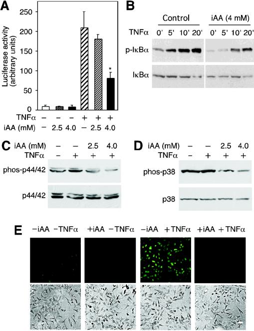
定点诱变。使用Stratagene的QuikChange XL定点诱变试剂盒按照制造商的说明进行定点诱变。 去:结果细胞内维生素C抑制TNF-α诱导的NF-κB活化。我们使用HeLa细胞作为模型系统,研究了维生素C抑制NF-κB活化的分子机制。通过将细胞暴露于DHA,使其富含维生素C(6)。该程序规避了组织培养基中与AA的促氧化作用相关的伪影(7)。与我们先前的发现一致,用NF-κB依赖的荧光素酶报告基因构建体(pNFκB-luc)转染的HeLa细胞在用TNF-α处理后诱导了荧光素酶活性增加了20倍(图(图1A).1A)。 )。装有4.0 mM AA的细胞显示萤光素酶活性的诱导作用较弱,约为八倍,对应于萤光素酶活性的60%抑制。细胞内浓度为2.5 mM AA对TNF-α诱导的萤光素酶活性没有显着影响(P = 0.21,n = 3,单尾t检验)(图(图1A).1A)。 TNF-α诱导的IκBα磷酸化,而装有4 mM AA的细胞显示IκBα的磷酸化降低(图(图1B).1B)。这些结果表明,细胞内维生素C通过阻止IκBα的磷酸化而抑制TNF-α诱导的NF-κB转录反应。
图。 1。
细胞内维生素C(iAA)抑制TNF-α诱导的信号传导。 (A)将转染了装有维生素C的pNFκB-luc报告基因构建体的HeLa细胞与TNF-α(+)孵育6小时,然后测定荧光素酶活性。数据表示为三个样本的平均值±标准差,星号表示装载维生素C的细胞与未装载维生素C的细胞之间的统计学差异(P = 0.001,n = 3;单尾学生t检验) 。 (B)将装载有4mM AA或未装载AA的细胞与TNF-α一起温育印迹上指示的时间(分钟)。制备细胞提取物,并通过抗磷酸化-IκBα抗体的免疫印迹法观察磷酸化的IκBα(p-IκBα)。未磷酸化的IκBα显示在下面的印迹中。 (C)用TNF-α处理载有维生素C(iAA)的HeLa细胞10分钟,并通过免疫印迹检测磷酸化(phos-p44 / 42)和非磷酸化p44 / 42 MAPK(p44 / 42)。 (D)用TNF-α处理装有AA的细胞,并显示了磷酸化(phos-p38)和非磷酸化p38 MAPK(p38)的免疫印迹。 (E)将载有4 mM细胞内维生素C(+ iAA)或未载有4 mM细胞内维生素C(-iAA)的HeLa细胞用TNF-α(+TNFα)处理,并用DCF通过荧光显微镜测定ROS(顶部面板)。显示了用光学显微镜观察的细胞(下图)。TNF-α诱导MAPK途径激活p44 / 42 MAPK和p38 MAPK(16)。因此,我们调查了细胞内AA是否也可以抑制这些激酶的激活。通过免疫印迹法检测,TNF-α诱导HeLa细胞中p44 / 42 MAPK磷酸化(图(图1C).1C)。用AA加载细胞可抑制TNF-α诱导的p44 / 42 MAPK磷酸化(图。(图1C).1C)。用TNF-α处理的细胞显示磷酸化的p38 MAPK适度增加,但用AA加载细胞导致p38 MAPK磷酸化显着降低(图。(图1D).1D)。由于TNF-α激活NF-κB涉及ROS(11),因此我们测量了维生素C负载的细胞中TNF-α诱导的ROS的相对水平。使用DCF和荧光显微镜检测细胞内ROS。 TNF-α治疗可引起细胞内ROS增加;然而,几乎没有检测到装有4 mM维生素C的细胞中的ROS水平(图(图1D).1D)。这些结果表明,维生素C通过淬灭参与IKK活化的ROS来抑制IκBα的磷酸化,从而通过抑制IκBα的磷酸化而阻止了TNF-α依赖性的NF-κB的活化,并且还降低了信号传导所引起的ROS的积聚。
细胞内维生素C抑制组成型活性IKKβ。
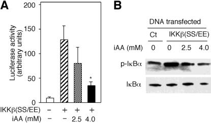
IKKβ被丝氨酸177和181处的磷酸化激活,并使IκBα磷酸化。用谷氨酸替代这些丝氨酸可产生组成性活性的IKKβ,IKKβ(SS / EE)(38)。为了研究维生素C除抑制IKKβ活化外还有另一种作用方式的可能性,我们分析了装有维生素C的细胞中IKKβ(SS / EE)的活性。pNFκB-luc和表达IKKβ(SS / EE)导致萤光素酶活性增加了12倍(图(图2A).2A)。然而,载有4 mM维生素C的细胞抑制了IKKβ(SS / EE)依赖的荧光素酶活性。较低浓度的AA(2.5 mM)没有显着影响(P = 0.11,n = 3,单尾t检验)(图(图2A).2A)。为了确定维生素C是否抑制IKKβ(SS / EE)的活性,我们测量了用IKKβ(SS / EE)转染的细胞中磷酸化IκBα的水平。在装有维生素C的细胞中,IKKβ(SS / EE)依赖性的IκBα磷酸化受到抑制(图(图2B).2B)。对IKKβ(SS / EE)的分析表明,细胞内维生素C对其表达没有影响(数据未显示)。因此,在载有维生素的细胞中,由IKKβ(SS / EE)的表达诱导的对IκBα磷酸化的抑制作用直接指示了维生素C对IKKβ(SS / EE)活性的抑制。
图。 2。
细胞内维生素C(iAA)抑制组成型活性IKKβ。 (A)用指示浓度的AA加载用pNFκB-luc共转染的HeLa细胞和编码组成型活性IKKβ[IKKβ(SS / EE)]的质粒。荧光素酶活性以任意单位测量,并表示为三个样品的平均值±标准偏差。星号表示装载了维生素C的细胞与未装载维生素C的细胞之间的统计学差异(P = 0.006,n = 3;单尾学生t检验)。 (B)将用IKKβ(SS / EE)表达载体或对照载体(Ct)转染的HeLa细胞加载2.5或4mMAAA。显示了磷酸化(p-IκBα)和总IκBα的免疫印迹。
维生素C的氧化形式DHA在体外抑制IKKβ和IKKα的活性。
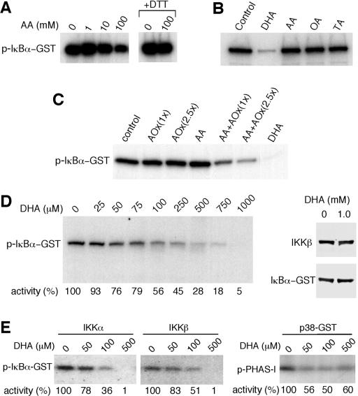
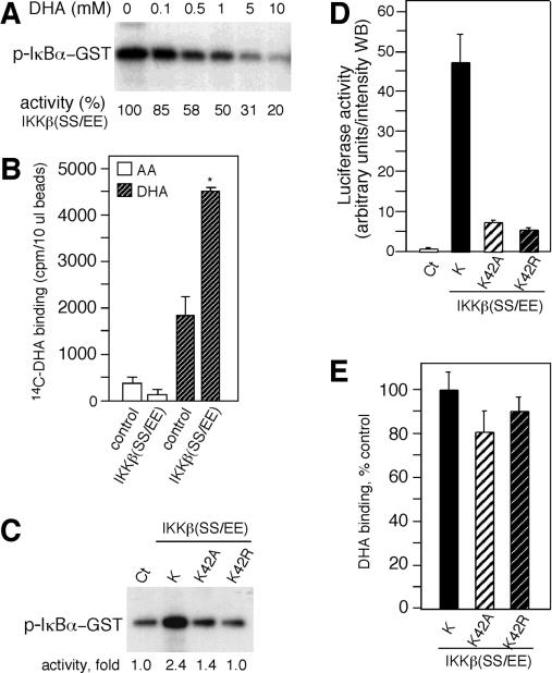
由于细胞内维生素C抑制了IKKβ(SS / EE)诱导的IκBα磷酸化,因此我们研究了AA是否会直接抑制无细胞系统中IKKβ的激酶活性。通过免疫沉淀从TNF-α处理的HeLa细胞中分离得到的活化的IKKβ被固定在蛋白G-琼脂糖珠上,然后进行体外激酶活性的测定(6)。向激酶反应混合物中添加1或10 mM AA对IKKβ活性没有影响(图(图3A).3A)。在相似的测定条件下,100 mM的AA抑制了IKKβ活性,但是当激酶反应混合物中包含二硫苏糖醇(DTT)时,这种抑制作用就消失了,表明该抑制活性与AA的氧化有关(图。(图3A).3A )。在非还原条件下,AA氧化为DHA,然后可以水解为草酸和苏氨酸(13,14)。
图。 3。
维生素C(DHA)的氧化形式在体外抑制IKKβ和IKKα。 (A)测定了用抗IKKβ抗体从TNF-α处理的HeLa细胞提取物中免疫沉淀的IKKβ的IKKβ活性。如所示,将激酶反应混合物与AA一起孵育。在某些情况下(+ DTT)将DTT添加到激酶反应混合物中。通过放射自显影观察磷酸化的GST-IkBα(p-IκBα-GST)。 (B)在缓冲液(对照)或1mM DHA,AA,草酸(OA)或苏糖酸(TA)存在下测定IKKβ活性。 (C)在缓冲液,抗坏血酸氧化酶(AOx),AA,酶促产生的DHA(AA + AOx)或DHA的存在下测定IKKβ活性。分别使用了一百或250 U抗坏血酸氧化酶[AOx(1x)或AOx(2.5x)。 (D)如印迹上所示,在不同浓度的DHA存在下测定IKKβ活性,相对激酶活性表示为印迹以下对照的百分比。通过用抗IκBα抗体或抗IKKβ抗体进行免疫印迹,可以观察到与缓冲液或1 mM DHA孵育30分钟的激酶反应混合物中的GST-IκBα和IKKβ。 (E)体外测定从转染细胞免疫沉淀的活化的IKKα和IKKβ的激酶活性。在DHA存在下体外测定p38 MAPK(p38-GST)的激酶活性。 PHAS-1被用作底物(p-PHAS-1)。通过放射自显影确定磷酸化的蛋白质,并且相对激酶活性显示为印迹以下的百分比。
为了确定AA的这些衍生物是否可以抑制IKKβ的活性,我们将激酶反应混合物与1 mM AA,DHA,草酸或苏糖酸一起孵育。与1 mM DHA一起孵育抑制了IKKβ的体外激酶活性(图(图3B).3B)。相反,将激酶反应混合物与1 mM AA,草酸或苏糖酸一起孵育时,未检测到对IKKβ活性的影响(图(图3B).3B)。为排除DHA制剂中激酶抑制性污染物的可能性,使用抗坏血酸氧化酶从AA酶促生产DHA,并测定其抑制活性。酶促衍生自AA的DHA也抑制IKKβ活性。单独的AA和抗坏血酸氧化酶没有抑制作用,证实了DHA的活性(图。(图3C).3C)。我们发现抑制大约50%的IKKβ激酶活性需要大约100μMDHA(图(图3D).3D)。 DHA的抑制活性不受DTT的影响(数据未显示)。用1 mM DHA孵育30分钟不会引起底物或IKKβ的降解(图(图3D),3D),证实DHA在体外直接抑制了IKKβ的酶活性。我们还研究了DHA是否抑制IKKα和p38 MAPK的活性。从转染细胞中分离的IKKα和IKKβ的激酶活性被DHA抑制。在500μM下,DHA几乎抑制了所有激酶活性(图。(图3E).3E)。 p38 MAPK也被DHA抑制,但500μMDHA抑制了约50%的激酶活性(图。(图3E3E)。
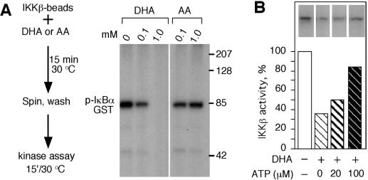
为了进一步研究DHA抑制激酶的机理,将固定在蛋白G上的内源性IKKβ与DHA或AA一起温育,并在激酶反应之前将磁珠洗净以去除AA和DHA(图。(图4A).4A) 。与1 mM DHA预孵育的IKKβ被完全抑制,而与0.1 mM DHA预孵育的保留率约为40%。用AA预孵育不会导致IKKβ活性降低(图(图4A).4A)。这些发现表明,DHA可能通过改变其酶促性质来抑制IKKβ,可能是直接结合激酶并干扰ATP或底物结合或催化。洗珠后,DHA抑制作用得以维持,表明DHA不与ATP或底物结合。通过分析在ATP存在下孵育DHA是否会抑制IKKβ活性来研究对ATP结合的干扰。 ATP部分竞争DHA的抑制活性(图(图4B),4B),这暗示DHA可能会干扰ATP与IKKβ的结合。
图。 4。
ATP竞争DHA的抑制活性。 (A,左)该过程的示意图。 15分钟15分钟(右)按照图3,3的说明通过放射自显影确定IKKβ的激酶活性,并显示磷酸化的IκBα-GST的免疫印迹。 (B)在缓冲液或20或100μMATP的存在下,将IKKβ珠与DHA孵育。激酶活性显示为对照的百分比。插入片段显示了磷酸化的GST-IκBα(pIκBα-GST)。
在体外,DHA抑制并结合组成型活性IKKβ(SS / EE)。
为了研究DHA是否结合IKKβ,我们使用了从转染细胞中分离的IKKβ(SS / EE)。在结合研究之前,我们确定DHA是否在体外抑制IKKβ(SS / EE)活性。剂量反应抑制试验表明1 mM DHA抑制了IKKβ(SS / EE)的活性40%(图(图5A).5A)。接下来,我们研究了维生素C是否与IKKβ(SS / EE)结合。将含有IKKβ(SS / EE)的珠子与[14C] DHA或[14C] AA孵育。与DHA的结合活性相比,AA显示与对照提取物或含有IKKβ(SS / EE)的珠预孵育的珠结合力低(图。(图5B).5B)。 DHA与IKKβ珠的结合比与对照提取物预温育的珠的结合大两倍(图(图5B).5B)。由于ATP降低了DHA的抑制活性,因此我们确定DHA是否可以与IKKβ(SS / EE)上突变的ATP结合位点结合。在所有激酶的ATP结合位点中发现的恒定赖氨酸突变会导致ATP结合丧失,进而导致激酶活性丧失(15)。产生了在IKKβ(SS / EE)中具有赖氨酸42的突变突变体,变为丙氨酸(K42A)或精氨酸(K42R),并在体外测定了激酶活性。两种突变体的激酶活性均存在缺陷(图。(图5C).5C)。与这些结果一致的是,IKKβ(SS / EE)突变体的转染显示萤光素酶活性降低(图。(图5D).5D)。 DHA以与IKKβ(SS / EE)相同的水平结合到IKKβ(SS / EE)K42R和K42A(P = 0.14,n = 3单尾t检验)(图(图3E).3E) 。这些结果表明DHA与IKKβ的结合独立于功能性ATP结合位点,并暗示了与ATP结合口袋不同的离散结合位点。
图。 5,
DHA与IKKβ结合。 (A)在体外测定从转染细胞中免疫沉淀的组成型活性IKKβ,IKKβ(SS / EE)的激酶活性。通过放射自显影确定激酶活性。 (B)将与抗FLAG珠结合的IKKβ(SS / EE)与[14C] DHA和[14C] AA一起温育,并用FLAG肽洗脱,并通过闪烁光谱法测定与IKKβ(SS / EE)有关的放射性。使用与未转染细胞(对照)提取物一起孵育的珠子,可以估计与抗FLAG珠子非特异性相关的放射性。 (C)在体外测定IKKβ(SS / EE)和ATP结合位点突变体IKKβ(SS / EE)K42A和K42R的激酶活性。量化数字化图像,激酶活性显示为印迹以下的增加倍数。 (D)用pNFκB-luc报告基因构建体和IKKβ(SS / EE)(K),K42A和K42R共转染的HeLa细胞的萤光素酶活性被测定。萤光素酶活性以Western印迹(WB)强度的任意单位显示。从用空载体共转染的细胞获得对照提取物(Ct)。 (E)将含有IKKβ(SS / EE)(K),K42A和K42R的珠子与[14C] DHA一起孵育,其结合活性显示为与IKKβ(SS / EE)的对照结合的百分比。
氧化应激将AA转化为DHA抑制了IKKβ(SS / EE)诱导的萤光素酶活性。
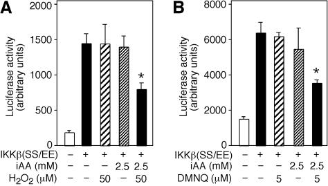
为了通过氧化应激源(例如H2O2)从AA生成细胞内DHA,我们首先确定了对细胞活力或荧光素酶活性没有影响的H2O2浓度。用IKKβ(SS / EE)转染并用50μMH2O2处理或未用H2O2处理的细胞显示相同的萤光素酶活性。但是,装有2.5 mM AA并与50μMH2O2孵育的细胞显示萤光素酶活性降低了约50%(P = 0.004,n = 3,单尾t检验)(图。(图6A).6A )。单独的细胞内AA(2.5 mM)没有显着影响(P = 0.5,n = 3,单尾t检验)。
图。 6。
细胞内DHA抑制IKKβ(SS / EE)诱导的萤光素酶活性。 (A)用pNFκB-luc报告基因构建体和IKKβ(SS / EE)(+)共转染的细胞装有2.5 mM iAA(细胞内维生素C)或未装有iAA(-)并用50μMH2O2处理或未用H2O2(-)。制备细胞提取物,测定萤光素酶活性并以任意单位表达。 (B)用5μMDMNQ处理或未用DMNQ(-)处理转染了pNFκB-luc和IKKβ(SS / EE)(+)的2.5mM iAA或未装载iAA(-)的细胞共转染的细胞。 4小时后,制备细胞提取物,荧光素酶活性表示为任意单位。
我们调查了ROS的细胞内生成器,例如二甲氧萘并萘醌(DMNQ),是否还会抑制预加载了2.5 mM维生素C的细胞中IKKβ(SS / EE)依赖的萤光素酶活性。DMNQ的浓度对萤光素酶活性或发现用IKKβ(SS / EE)转染的细胞的生存力为5μM。装有2.5 mM维生素C并与5μMDMNQ孵育的细胞显示萤光素酶活性显着降低(P = 0.003,n = 3,单尾t检验)(图(图6B).6B)。这些结果表明,AA向DHA的细胞内转化抑制了IKKβ(SS / EE)的激酶活性,表明DHA在体内起激酶抑制剂的作用。我们建议,在装有维生素C并置于氧化条件下的细胞中,AA可以抑制ROS并转化为DHA。生成的DHA可以通过促进性葡萄糖转运蛋白离开细胞,或作为直接激酶抑制剂发挥作用,如此处所示,用于IKKα和IKKβ(图(图7).7)。这些过程通过抗氧化剂维生素C将氧化应激与激酶抑制联系起来。
图。 7。
维生素C调节信号响应的示意图。维生素C作为DHA通过葡萄糖转运蛋白进入细胞,并迅速还原为AA。 ROS通过激活IKKβ诱导NF-κB信号传导应答,而AA猝灭ROS,抑制IKKβ的激活。在这些过程中,AA氧化成DHA,而DHA抑制IKKβ。
讨论
ROS是在细胞代谢过程中产生的,并且也作为信号转导途径的重要介体参与。因此,抗氧化剂可能在调节细胞对外部刺激的反应中起关键作用(24)。 ROS的代谢积累与局部受体激活诱导的ROS之间的区别定义了细胞内维生素C在信号转导中的特殊作用(5、27)。例如,已知粒细胞-巨噬细胞集落刺激因子(GM-CSF)和白介素3信号涉及ROS(5,30),维生素C在Jak2激活水平抑制GM-CSF反应,抑制β磷酸化GM-CSF受体(5)。还已知NF-κB应答在信号转导中利用ROS,并且已经表明,NF-κB的激活被抗氧化剂酶和抗氧化剂例如吡咯烷二硫代氨基甲酸酯,N-乙酰基-1-半胱氨酸,硫氧还蛋白,谷胱甘肽的过量表达抑制。和维生素C(4、6、9、11、23)。在这项研究中,我们试图以机械方式定义维生素C在抑制IκBα磷酸化中的作用。
维生素C作为细胞内抗氧化剂的重要性已得到充分证明,并能抑制过氧化氢诱导的细胞死亡(12、37),FAS结合CD95诱导的细胞凋亡(27)和氧化应激诱导的DNA损伤(22)。除了这些功能外,维生素C是各种酶促羟基化反应的辅助因子(13、14)。维生素C的所有这些功能,包括其作为辅因子的功能,都依赖于其提供电子,在此过程中被氧化成DHA的能力。 DHA主要在维生素C的运输中起作用(36),但据报道也抑制了己糖激酶活性(8a)。
我们发现DHA可以充当激酶抑制剂,并直接抑制IKKβ,IKKα和p38 MAPK的活性,从而扩展了我们对维生素C如何调节ROS依赖性NF-κB信号传导的认识(4、6)。维生素C抑制IKKβ酶活性的最初证据是由组成型活性IKKβ[IKKβ(SS / EE)]诱导的反应的下调。体外研究表明,维生素C的氧化形式抑制了免疫纯化的IKKβ和IKKβ(SS / EE)。使用放射性标记的[14C] DHA,我们发现DHA与IKKβ结合,而AA在体外既不显示抑制活性,也不显示与IKKβ的结合活性。DHA对IKKα和β酶活性的抑制作用定义了两种由维生素C负调节NF-κB反应的机制。首先,AA抑制由细胞因子-受体相互作用诱导的ROS信号传导,从而阻止ROS介导的反应的激活,并淬灭由信号产生的ROS。其次,由于AA氧化,会生成DHA,并抑制ROS激活的激酶。在载有AA的细胞中通过实验证明了这一点,并通过用H2O2或DMNQ对其施加压力来诱导生成DHA。这些发现表明维生素C在抑制IKKβ引起的TNF-α信号启动和转导方面具有抗炎作用。
DHA抑制激酶的确切机制尚不确定,但我们建议DHA结合至激酶活性位点附近的口袋。 ATP初始竞争实验的结果表明,它可以与ATP结合位点相互作用,但是结合测定的结果表明,完整的ATP位点对于DHA结合不是必需的。这些明显的矛盾结果可以通过存在独立的DHA结合位点来调节。对接模拟和计算机建模表明,DHA可以在催化位点附近结合而不在ATP结合位点结合(数据未显示)。
据报道,维生素C抑制兔肌肉腺苷酸激酶的酶活性。但是,首先将AA与DTT孵育即可克服这种抑制作用(29)。这些结果表明,AA被转化为DHA,而DHA可能是抑制腺苷酸激酶的分子。据报道,可溶性胍基环化酶也被AA抑制(31)。加载了维生素C并处于氧化应激状态的细胞在G2 / M检查点诱导了短暂的细胞周期停滞,并且推测DHA通过延迟细胞周期蛋白B-cdc2的活化来诱导细胞周期停滞(3)。可以通过假设DHA可能影响在细胞周期控制中重要的激酶反应来使这些结果合理化。我们的数据表明DHA可以抑制IKKβ,IKKα和p38 MAPK,而T4多核苷酸激酶对DHA抑制有抵抗力(数据未显示)。
NF-κB活化抑制剂,例如水杨酸盐和阿司匹林,在减少炎症和可能的肿瘤发生中起重要作用。这些药物通过与ATP结合竞争来抑制IKKβ的激酶活性(38)。我们将维生素C作为NF-κB抑制剂的发现表明,维生素C在炎症和细胞凋亡中发挥了作用。维生素C可以通过两种不同但相关的机制来抑制NF-κB的激活:下调ROS诱导的激活并直接抑制IKKα和IKKβ。Vitamin C Is a Kinase Inhibitor: Dehydroascorbic Acid ...
https://www.ncbi.nlm.nih.gov/pmc/articles/PMC444845
Our findings with vitamin C as an NF-κB inhibitor point to a role for the vitamin in inflammation and apoptosis. Vitamin C can inhibit the activation of NF-κB by two distinct but related mechanisms: down-regulating ROS induced activation and directly inhibiting IKKα and IKKβ.
Cited by: 106
Publish Year: 2004
Author: JuDepartment of Clinical Laboratories,1 Program in Molecular Pharmacology and Chemistry, Memorial Sloan-Kettering Cancer Center,2 Structural Biology Program, Department of Physiology and Biophysics, Mount Sinai School of Medicine, New York, New York3
*Corresponding author. Mailing address: Program in Molecular Pharmacology and Chemistry, Box 451, Memorial Sloan-Kettering Cancer Center, New York, NY 10021. Phone: (212) 639-8483. Fax: (212) 772-8589. E-mail: gro.ccksm.iks@edlog-d.
维生素C通过抑制IκBα磷酸化抑制TNFα诱导的NFκB活化,影响炎症、肿瘤和凋亡过程
Vitamin C Suppresses TNFα-Induced NFκB Activation by Inhibiting IκBα Phosphorylation†
摘要
细胞外刺激信号激活转录因子NFκB,导致涉及免疫应答,炎症和细胞存活的基因表达调节过程。肿瘤坏死因子α(TNFα)通过涉及NFκB诱导激酶(NIK),其激活下游多亚基IκB激酶(IKK)明确定义的激酶途径激活NFκB。 IKK依次磷酸化IκB(NFκB功能的中央调节剂)。我们发现细胞内维生素C以剂量依赖性方式抑制人细胞系(HeLa,单核U937,髓样白血病HL-60和乳腺MCF7)和原代内皮细胞(HUVEC)中TNFα诱导的NFκB活化。维生素C是一种重要的抗氧化剂,大多数细胞通过输送氧化形式的维生素C-脱氢抗坏血酸(DHA)在细胞内积累抗坏血酸(AA)。因为抗坏血酸在体外存在过渡金属的情况下是强氧化剂,所以我们通过将细胞与DHA一起孵育为细胞加载了维生素C。装有维生素C的细胞显示出TNFα诱导的NFκB核易位,NFκB依赖的报告基因转录和IκBα磷酸化显着降低。我们的数据指出了维生素C通过抑制TNFα诱导的独立于p38 MAP激酶的NIK和IKKβ激酶激活来抑制NFκB激活的机制。这些结果表明,细胞内的维生素C可以通过抑制NFκB活化的影响炎症,肿瘤,和凋亡过程。
Extracellular stimuli signal for activation of the transcription factor NFκB, leading to gene expression regulating processes involved in immune responses, inflammation, and cell survival. Tumor necrosis factor-α (TNFα) activates NFκB via a well-defined kinase pathways involving NFκB-inducing kinase (NIK), which activates downstream multisubunit IκB kinases (IKK). IKK in turn phosphorylates IκB, the central regulator of NFκB function. We found that intracellular vitamin C inhibits TNFα-induced activation of NFκB in human cell lines (HeLa, monocytic U937, myeloid leukemia HL-60, and breast MCF7) and primary endothelial cells (HUVEC) in a dose-dependent manner. Vitamin C is an important antioxidant, and most cells accumulate ascorbic acid (AA) intracellularly by transporting the oxidized form of the vitamin, dehydroascorbic acid (DHA). Because ascorbic acid is a strong pro-oxidant in the presence of transition metals in vitro, we loaded cells with vitamin C by incubating them with DHA. Vitamin C-loaded cells showed significantly decreased TNFα-induced nuclear translocation of NFκB, NFκB-dependent reporter transcription, and IκBα phosphorylation. Our data point to a mechanism of vitamin C suppression of NFκB activation by inhibiting TNFα-induced activation of NIK and IKKβ kinases independent of p38 MAP kinase. These results suggest that intracellular vitamin C can influence inflammatory, neoplastic, and apoptotic processes via inhibition of NFκB activation.
Cited by: 259
Publish Year: 2002
Author: Juan M. Cárcamo, Alicia Pedraza, Oriana BóProgram in Molecular Pharmacology and Chemistry and Departments of Clinical Chemistry and Medicine, Memorial Sloan-Kettering Cancer Center, 1275 York Avenue, Box 451, New York, New York 10021
PPARalpha对细胞代谢和炎症的抗癌特性。底线:以前,PPAR配体与糖尿病,高脂血症和心血管疾病有关,因为它们调节调节葡萄糖和脂质代谢的基因的表达.PPARalpha配体的抗增殖,促凋亡,抗炎和潜在的抗转移特性的新兴证据提示我们讨论PPARalpha在肿瘤抑制中的可能作用。在有限的养分利用率的状态下(经常存在于肿瘤微环境中),PPARalpha与AMP依赖的蛋白激酶(AMPK)协同作用:(i)抑制致癌Akt活性,(ii)抑制细胞增殖(iii)迫使糖酵解依赖性癌细胞陷入“代谢灾难 Metabolic catastrophy”。 PPARalpha的其他潜在抗癌作用包括抑制炎症和上偶联蛋白(UCP)的上调,从而减弱线粒体活性氧的产生和细胞增殖。
摘要
过氧化物酶体增殖物激活受体(PPARs)作为治疗靶点最近引起了广泛的关注。以前,PPAR配体与糖尿病,高脂血症和心血管疾病有关,因为它们可调节调节葡萄糖和脂质代谢的基因的表达。最近,PPAR配体也被认为是潜在的抗癌药,具有较低的全身毒性。 PPARalpha配体的抗增殖,促凋亡,抗炎和潜在的抗转移特性的新兴证据促使我们讨论PPARalpha在肿瘤抑制中的可能作用。 PPARalpha激活可通过阻断脂肪酸合成并促进脂肪酸β-氧化来靶向癌细胞的能量平衡。在有限的养分利用率的状态下(通常存在于肿瘤微环境中),PPARalpha与AMP依赖的蛋白激酶(AMPK)协同作用:(i)抑制致癌的Akt活性,(ii)抑制细胞增殖,以及(iii)强迫糖酵解依赖性癌细胞陷入“代谢灾难”。 PPARalpha的其他潜在抗癌作用包括抑制炎症和上偶联蛋白(UCP)的上调,从而减弱线粒体活性氧的产生和细胞增殖。
总之,有很强的前提,即低毒性和耐受性良好的PPAR配体应被视为对抗弥散性癌症的新治疗剂,这代表了现代医学和基础研究的主要挑战。
图3:PPARα通过抑制主要的炎性转录因子NFκB和AP-1拮抗主要的炎性信号通路。此外,PPARα通过上调解偶联蛋白UCP2和UCP3减少ROS介导的炎症。有关更多详细说明,请参见文本。箭头表示激活/上调,而虚线表示细胞蛋白或过程的抑制/下调。 Erk1 / 2-细胞外信号应答激酶1/2; IκB-NFκB抑制剂; MAPK-促分裂原活化蛋白激酶; NFκB—核因子κB; ROS-活性氧。
Fig3: PPARα antagonizes main inflammatory signaling pathways throughrepression of the main inflammatory transcription factors: NFκB and AP-1.Additionally, PPARα reduces ROS-mediated inflammation by upregulation ofuncoupling proteins UCP2 and UCP3. See the text for more detailedexplanation. Arrowheads represent activation/upregulation, and blunted linesindicate inhibition/downregulation of the cellular proteins or processes.AP-1—activating protein-1; Erk1/2—extracellular signal response kinase 1/2; IκB—inhibitor of NFκB; MAPK—mitogen activated protein kinase; NFκB—nuclear factor κB; ROS—reactive oxygen species.
Affiliation: Department of Food Biotechnology, Faculty of Food Technology, Agricultural University of Krakow, ul. Balicka 122, 31149 Krakow, Poland.
EPA和DHA降低HK-2细胞中LPS诱导的炎症反应:PPAR-γ依赖性机制的证据
EPA and DHA reduce LPS-induced inflammation responses in HK-2 cells: Evidence for a PPAR-γ–dependent mechanism
背景
最近的研究表明,鱼油中含有ω-3多不饱和脂肪酸(ω-3PUFAs)二十碳五烯酸(EPA)(C20:5ω3)和二十二碳六烯酸(DHA)(C22:6ω3)会延缓进程肾脏疾病的发生,特别是在IgA肾病(IgAN)中。尽管人们越来越了解鱼油的有益作用,但对ω-3PUFA的作用机理知之甚少。据报道,过氧化物酶体增殖物激活受体(PPAR)的激活抑制了促炎细胞因子的产生。 EPA和DHA均显示可激活PPAR。这项研究的目的是检查ω-3PUFA是否通过激活人肾小管细胞中的PPARs具有抗炎作用。
方法
在所有实验中均使用了永生化的人近端肾小管细胞系[人肾2(HK-2)细胞]。从ω-3PUFA处理过的细胞中收集条件培养基,并进行酶联免疫吸附测定(ELISA)。从上述细胞中分离出总细胞RNA,用于实时定量聚合酶链反应(PCR)。从HK-2细胞制备核提取物用于转录因子激活测定。
结果
EPA和DHA浓度分别为10μmol/ L和100μmol/ L均有效降低了脂多糖(LPS)诱导的核因子-κB(NF-κB)活化和单核细胞趋化蛋白1(MCP-1)的表达。 EPA和DHA还能增加HK-2细胞中PPAR-γmRNA和蛋白质活性(2到3倍)。剂量为100μmol/ L的双酚A二缩水甘油醚(BADGE)消除了EPA和DHA诱导的PPAR-γ活化,并消除了EPA和DHA对LPS诱导的HK-2细胞中NF-κB活化的抑制作用。与存在EPA和DHA的对照细胞相比,PPAR-γ的过表达进一步抑制了NF-κB的活化。
结论
我们的数据表明,EPA和DHA均可通过HK-2细胞中PPAR-γ依赖性途径下调LPS诱导的NF-κB活化。这些结果表明,EPA和DHA激活PPAR-γ可能是鱼油有益作用的潜在机制之一。EPA and DHA reduce LPS-induced inflammation responses in HK-2 cells: Evidence for a PPAR-γ–dependent mechanism - ScienceDirect
https://www.sciencedirect.com/science/article/pii/S0085253815505322
The Inflammation Link: NF-κB Remains a Difficult but Intriguing Target
While anticancer therapies aimed at particular pathways have mushroomed in recent years, one crucial target has remained elusive: nuclear factor kappa-light-chain-enhancer of activated B cells (NF-κB). The pathway is constitutively active in the majority of cancers and provides a mechanistic link between chronic inflammation and tumorigenesis. As such, it represents an important target for anticancer therapy. The complex regulation of NF-κB activation has presented significant challenges for the development of such agents, but researchers are now beginning to better understand and embrace this complexity to drive development of a variety of novel NF-κB-targeting strategies.
The NF-κB Family
NF-κB was discovered in late 1980s as a nuclear factor that binds to the enhancer region of the kappa-light chain of immunoglobulin in B cells. It was subsequently shown to be a ubiquitous family of transcription factors that are present in all cells and control the transcription of more than 500 target genes involved in critical cellular pathways including proliferation and apoptosis. The NF-κB pathway is activated by a wide range of stimuli, including stress, cytokines, free radicals, ultraviolet irradiation, and bacterial or viral antigens.
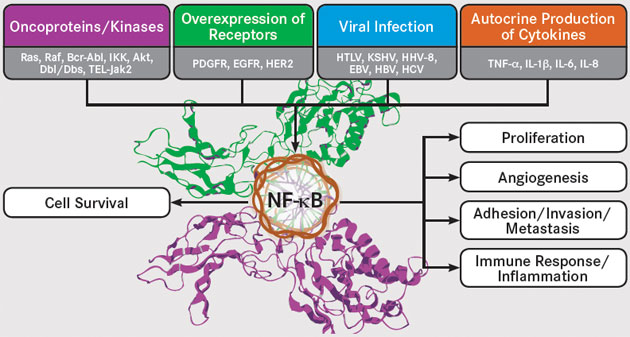
NF-κB is activated by a number of upstream signaling pathways, including the epidermal growth factor receptor (EGFR) and human epidermal growth factor receptor-2 (HER2), platelet-derived growth factor receptor (PDGFR), and c-KIT pathways. Another key activator of NF-κB is the receptor-activator of NF-κB (RANK), which is a type of tumor necrosis factor (TNF) receptor. Further adding to the complex regulation of NF-κB signaling, it undergoes numerous posttranslational modifications, including methylation and acetylation, and it interacts with several other transcription factors, such as signal transducer and activator of transcription (STAT)-3 and hypoxia inducible factor (HIF)-1α, both of which regulate NF-κB activity.
The Many Roles of NF-κB
Roles of NF-kB EBV, indicates Epstein-Barr virus; EGFR, epidermal growth factor receptor; HER2, human epidermal growth factor receptor 2; HBV, hepatitis B virus; HCV, hepatitis C virus; HHV-8, human herpesvirus 8; HTLV, human T-lymphotropic virus; IL, interleukin; PDGFR, platelet-derived growth factor receptor; TNF, tumor necrosis factor. Adapted from Sethi G, Sung B, Aggarwal BB. Nuclear factor-κB activation: from bench to bedside. Exp Biol Med. 2008; 233(1): 21-31.
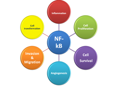
Linking Inflammation and Cancer The activation of NF-κB plays a role in the suppression of apoptosis, stimulation of proliferation, and promotion of migration and invasion, all of which are hallmarks of cancer. Unsurprisingly, therefore, NF-κB has been implicated in tumorigenesis. What is surprising is that oncogenic mutations in the NF-κB genes are rare and largely limited to lymphoid malignancies, yet NF-κB activation is observed in almost all tumors.
... to read the full story
炎症链接:NF-κB仍然是一个困难但有趣的目标
The Inflammation Link: NF-κB Remains a Difficult but Intriguing Target
图示:EBV表示爱泼斯坦-巴尔病毒; EGFR,表皮生长因子受体; HER2,人表皮生长因子受体2; HBV,乙肝病毒; HCV,丙型肝炎病毒; HHV-8,人类疱疹病毒8; HTLV,人类T淋巴病毒; IL,白介素; PDGFR,血小板衍生的生长因子受体; TNF,肿瘤坏死因子。
尽管近年来针对特定途径的抗癌疗法如雨后春笋般涌现,但一个关键的目标仍然难以捉摸:活化的B细胞核因子κ-轻链增强剂(NF-κB)。该途径在大多数癌症中具有组成性活性,并提供了慢性炎症和肿瘤发生之间的机制联系。因此,它代表了抗癌治疗的重要目标。 NF-κB激活的复杂调控为此类药物的开发提出了重大挑战,但是研究人员现在开始更好地理解和接受这种复杂性,以推动各种新型NF-κB靶向策略的发展。
NF-κB家族
NF-κB在1980年代末被发现是一种核因子,与B细胞中免疫球蛋白的κ轻链的增强子区域结合。随后证明它是一个存在于所有细胞中的普遍存在的转录因子家族,可控制涉及关键细胞途径(包括增殖和凋亡)的500多个靶基因的转录。 NF-κB途径被多种刺激激活,包括应激,细胞因子,自由基,紫外线照射以及细菌或病毒抗原。
NF-κB被许多上游信号通路激活,包括表皮生长因子受体(EGFR)和人表皮生长因子受体2(HER2),血小板源性生长因子受体(PDGFR)和c-KIT途径。 NF-κB的另一个关键激活剂是NF-κB受体激活剂(RANK),这是一种肿瘤坏死因子(TNF)受体。进一步增加了NF-κB信号的复杂调控,它经历了许多翻译后修饰,包括甲基化和乙酰化,并且与其他几种转录因子(例如信号转导子和转录激活子(STAT)-3和低氧诱导因子)相互作用( HIF)-1α,两者均调节NF-κB活性。
NF-κB的许多作用 NF-kB EBV的作用表明爱泼斯坦-巴尔病毒; EGFR,表皮生长因子受体; HER2,人表皮生长因子受体2; HBV,乙肝病毒; HCV,丙型肝炎病毒; HHV-8,人类疱疹病毒8; HTLV,人类T淋巴病毒; IL,白介素; PDGFR,血小板衍生的生长因子受体; TNF,肿瘤坏死因子。改编自Aghiwal BB的Sethi G,SungB。核因子-κB激活:从工作台到床边。 Exp Biol Med。 2008年; 233(1):21-31。 将炎症与癌症联系起来NF-κB的激活在抑制细胞凋亡,刺激增殖以及促进迁移和侵袭中发挥作用,所有这些都是癌症的标志。因此,毫不意外的是,NF-κB与肿瘤发生有关。令人惊讶的是,NF-κB基因中的致癌突变很少见,并且很大程度上限于淋巴样恶性肿瘤,但是几乎在所有肿瘤中都观察到了NF-κB激活。 ...阅读全文
The Inflammation Link: NF-κB Remains a Difficult but Intriguing Target
https://www.onclive.com/publications/oncology-live/2013/june-2013/the-inflammation-link-nf-b-remains-a-difficult-but-intriguing-target
炎症101:炎症助长癌症的燃料2012年11月11日,医学博士Brian D. Lawenda
我认为我们大多数人通常将炎症视为身体对伤害和感染的保护性反应。没有它,我们将陷入严重的困境(除非我们生活在一个密封的泡沫中。) 如果只是那么简单... 炎症分为两类,“急性”和“慢性”。
急性发炎:当我们的身体行为正常时,免疫细胞(白细胞,巨噬细胞等)在检测到组织中的氧化应激,损伤或感染时就会发挥作用。这些细胞向周围组织和血流中释放出大量的炎性化学物质和蛋白质,从而: 消灭任何外来入侵者(即细菌,病毒)向该区域发出更多的免疫细胞信号扩张血管以增加该区域的血流(使免疫细胞,氧气和营养物质流入)触发组织和新血管的生长(以帮助组织修复并向这些组织提供营养)此过程称为“急性”炎症,它将在感染得到控制或伤口治愈后的数小时至数周内解决……除非事实并非如此……
慢性炎症:
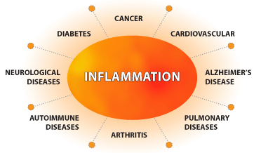
急性炎症可以保护我们免受敌对环境的侵害,而“慢性”炎症(长期处于低水平,闷热的炎症状态,持续数月至数年)会伤害甚至杀死我们。这种差异是现代医学最重要的发现之一。我们现在认识到,慢性炎症与几乎所有慢性疾病(包括癌症)的发生和发展有关。
慢性炎症的作用在自身免疫和慢性感染性疾病中尤为明显,在这些疾病中,涉及的组织充满了持续的化学和蛋白质信号传导,以修复和生长。
慢性炎症还会大大降低人体对胰岛素的敏感性,导致血糖水平持续升高,胰岛素和胰岛素样生长因子1(IGF-1)的产生增加,代谢综合征,2型糖尿病和肥胖症。所有这些因素均已显示会增加癌症发展,进展和复发的风险。
“炎性分子Inflammasome”
我们大多数人以前从未听说过“炎症小体”,因为它们是最近才发现的(2002年)。这些蛋白质复合物是人体免疫系统不可或缺的一部分。它们由特殊的“危险感应蛋白”组成,这些蛋白被病毒,细菌和自由基化学物质激活。
一旦被激活,炎症小体就会向我们的免疫细胞(巨噬细胞)发出信号,开始产生大量的炎症蛋白。最新研究表明,炎症小体在很大程度上归因于肥胖导致的慢性炎症状态。
核因子κB(NF-kB)在癌症中的作用
The Role of Nuclear Factor Kappa-B (NF-kB) in Cancer
当免疫细胞首次检测到任何组织损伤或压力的迹象时,它们就会激活一种称为核因子κB(NF-kB)的细胞蛋白。该蛋白是开启组织炎症的主要“开关”。活化的NF-kB信号通知细胞(通过许多途径,包括通过激活炎性小体)开始产生参与炎症和组织修复的多种蛋白质。
到目前为止,只要在癌前或癌变组织中没有出现生长信号,就可以了……
在慢性炎症条件下,发炎的组织比非发炎的组织更容易发生和积累DNA损伤。这是由于发炎的组织中大量自由基的产生(实际上,由于自由基的损伤和有毒的暴露,每天在我们的细胞中产生数十亿个DNA突变。)当这些高反应性化学物质与DNA相互作用时,它们可以产生DNA突变。只要细胞能够修复这些DNA突变(大多数细胞具有内置的DNA修复机制)或向细胞发出信号使其经历细胞自杀过程(“凋亡”),癌前细胞就不会发育。
但是,在持续暴露于NF-kB定向信号期间(在慢性炎症中发生),细胞凋亡和DNA修复机制均被关闭。这导致具有遗传突变的细胞积累。如果这些细胞产生足够的突变,它们可以转化为癌前细胞,最终转变为癌细胞。
但是,在持续暴露于NF-kB定向信号期间(在慢性炎症中发生),细胞凋亡和DNA修复机制均被关闭。这导致具有遗传突变的细胞积累。如果这些细胞产生足够的突变,它们可以转化为癌前细胞,最终转变为癌细胞。
在存在慢性炎症的情况下,癌前和癌性组织会发出信号,指示其分裂,生长甚至侵入周围组织(包括血液和淋巴管,从而使它们可以进入身体的其他部位);这被称为“转移性”扩散。)这些炎症信号还会使该区域的免疫细胞失活,从而使其无法识别和攻击新形成的癌细胞。
癌细胞学会确保自己的生长和存活的偷偷摸摸的事情之一是它们可以激活自己的NF-kB。这使他们能够打开周围组织中所有相同的炎症蛋白,从而劫持了人体的组织修复和生长机制。
实验表明,炎性小体(inflammasome)激活通过多步过程涉及NF-kB。当免疫细胞(巨噬细胞)识别出任何组织损伤或氧化应激时,它们首先会激活NF-kB。然后,活化的NF-kB向细胞发出信号以形成炎症小体蛋白复合物(步骤1和2),它们现已准备就绪(或“引发”)以进一步响应组织损伤或氧化应激。在此步骤(第3步)中,激活的炎症小体向细胞发出信号以产生和释放炎症蛋白(即IL-1等)。
Inflammation 101: The Fuel That Feeds Cancer
Nov 11, 2012 Brian D. Lawenda, M.D.
I think most of us typically think of inflammation as our body’s protective response to injury and infection. Without it, we’d be in serious trouble (unless we lived in a sealed bubble.)
If it were only that simple…
Inflammation comes in two categories, “acute” and “chronic.”
Acute Inflammation:
When our body is behaving properly, immune cells (white blood cells, macrophages, etc.) kick into gear when they detect oxidative stress, injury or infection in the tissues. These cells release a storm of inflammatory chemicals and proteins into the surrounding tissues and into the blood stream to:
Destroy any foreign invaders (i.e. bacteria, viruses)
Signal more immune cells to the area
Dilate blood vessels to increase blood flow to the area (to enable the influx of immune cells, oxygen and nutrients)
Trigger the growth of tissues and new blood vessels (to aid in tissue repair and supply nutrients to these tissues)
This process is called “acute” inflammation, and will resolve within hours-to-weeks after the infection is controlled or the injury is healed…unless it doesn’t…
Chronic Inflammation:
Whereas acute inflammation protects us from our hostile environment, “chronic” inflammation (a prolonged state of low-level, smoldering inflammation, lasting months-to-years) can hurt or even kill us. This difference is one of the most important discoveries of modern medicine. We now recognize that chronic inflammation is involved in the development and progression of almost every chronic disease…including cancer.
The role of chronic inflammation is particularly poignant in the autoimmune and chronic infectious diseases, where the involved tissues are flooded with persistent chemical and protein signaling to repair and grow.
Chronic inflammation can also significantly reduce your body’s sensitivity to insulin, leading to persistently elevated levels of blood sugar, increased production of insulin and insulin-like growth factor 1 (IGF-1), metabolic syndrome, type 2 diabetes and obesity. All of these factors have been shown to increase the risk of cancer development, progression and recurrence.
The “Inflammasome”
Most of us have never heard of “inflammasomes” before, as they were discovered relatively recently (in 2002.) These protein complexes are an integral part of our body’s immune system. They are comprised of special, ‘danger sensing proteins’ that are activated by viruses, bacteria, and free radical chemicals.
Once activated, the inflammasomes signal our immune cells (macrophages) to begin to produce a storm of inflammatory proteins. The latest research indicates that inflammasomes are responsible in large part for the chronic inflammatory state that results from obesity.The Role of Nuclear Factor Kappa-B (NF-kB) in Cancer
When immune cells first detect any signs of tissue injury or stress, they activate a cellular protein, called nuclear factor kappa-B (or NF-kB.) This protein is the main ‘switch’ that turns on the inflammation in the tissues. Activated NF-kB signals the cell (via many pathways, including through the activation of inflammasomes) to begin to produce numerous proteins involved in inflammation and tissue repair.
So far so good, as long as the growth signals are not happening in precancerous or cancerous tissues…
During chronic inflammatory conditions, inflamed tissues are predisposed to developing and accumulating DNA damage at a greater rate than non-inflamed tissues. This is due to the production of large amounts of free radicals in inflamed tissues [in fact, billions of DNA mutations develop in our cells each day as a result of free radical injury and toxic exposures.] When these highly-reactive chemicals interact with DNA, they can create DNA mutations. As long as the cell is able to either repair these DNA mutations (most cells have built-in DNA repair mechanisms) or signal the cell to undergo a process of cell suicide (“apoptosis”), precancerous cells will not develop.
However, during constant exposure to NF-kB directed signals (which happens during chronic inflammation), both apoptosis and DNA repair mechanisms are shut down. This leads to an accumulation of cells with genetic mutations. If these cells develop enough mutations, they can transform into precancerous cells and, eventually, cancer cells.
In the presence of chronic inflammation, precancerous and cancerous tissues are signaled to divide, grow and even invade into surrounding tissues (including blood and lymph vessels, which gives them access to the rest of the body; this is referred to as “metastatic” spread.) These inflammatory signals also inactivate immune cells in the area, preventing them from being able to identify and attack the newly formed cancer cells.
One of the sneaky things that cancer cells learn to do to ensure their own growth and survival, is that they can activate their own NF-kB. This enables them to turn on all of the same inflammatory proteins in the surrounding tissues, thereby hijacking the body’s tissue repair and growth mechanisms.
Experiments have shown that inflammasome activation involves NF-kB through a multi-step process. When the immune cells (macrophages) identify any tissue injury or oxidative stress, they first activate NF-kB. Activated NF-kB then signals the cells to form inflammasome protein complexes (steps 1 and 2), which are now ready (or ‘primed) to further respond to the tissue injury or oxidative stress. At this step (step 3), the activated inflammasome signals the cell to produce and release inflammatory protein (i.e. IL-1, etc.)Inflammation 101: The Fuel That Feeds Cancer - Integrative Oncology Essentials
https://integrativeoncology-essentials.com/2012/11/inflammation-101-the-fuel-that-feeds-cancer
Institute of General Pathology, 2Institute of Pathology, 3Institute of Pharmacology and 4Institute of Histology, Catholic University, L.go F. Vito, 1, 00168 Rome, Italy
Omega-3 PUFAs reduce VEGF expression in human colon cancer cells modulating the COX-2/PGE 2 induced ERK-1 and -2 and HIF-1α induction pathwayn-3 Polyunsaturated fatty acids (PUFAs) inhibit the development of microvessels in mammary tumors growing in mice. Human colorectal tumors produce vascular endothelial growth factor (VEGF) whose expression is up-regulated in tumor cells by both cyclooxygenase-2 (COX-2) and PGE 2 and directly correlated to neoangiogenesis and clinical outcome.
The goal of this study was to examine the capability of n-3 PUFAs to regulate VEGF expression in HT-29 human colorectal cells in vitro and in vivo . Constitutive VEGF expression was augmented in cultured HT-29 cells by serum starvation and the effects of eicosapentaenoic (EPA) or docosahexaenoic acid (DHA) on VEGF, COX-2, phosphorylated extracellular signal-regulated kinase (ERK)-1 and -2 and hypoxia-inducible-factor 1-α (HIF-1α) expression and PGE 2 levels were assessed.
Tumor growth, VEGF, COX and PGE 2 analysis were carried out in tumors derived from HT-29 cells transplanted in nude mice fed with either EPA or DHA. Both EPA and DHA reduced VEGF and COX-2 expression and PGE 2 levels in HT-29 cells cultured in vitro . Moreover, they inhibited ERK-1 and -2 phosphorylation and HIF-1α protein over-expression, critical steps in the PGE 2 -induced signaling pathway leading to the augmented expression of VEGF in colon cancer cells.
EPA always showed higher efficacy than DHA in vitro . Both fatty acids decreased the growth of the tumors obtained by inoculating HT-29 cells in nude mice, microvessel formation and the levels of VEGF, COX-2 and PGE 2 in tumors.
The data provide evidence that these n-3 PUFAs are able to inhibit VEGF expression in colon cancer cells and suggest that one possible mechanism involved may be the negative regulation of the COX-2/PGE 2 pathway. Their potential clinical application as anti-angiogenic compounds in colon cancer therapy is proposed.
n-3 PUFAs reduce VEGF expression in human colon cancer cells modulating the COX-2/PGE 2 induced ERK-1 and -2 and HIF-1α induction pathway | Carcinogenesis | Oxford Academic
https://academic.oup.com/carcin/article/25/12/2303/2475914
Omega-3脂肪酸抑制膀胱癌大鼠模型中的肿瘤生长
Omega-3 Fatty Acids Inhibit Tumor Growth in a Rat Model of Bladder Cancer
摘要
Omega-3(ω-3)脂肪酸已在多种癌症类型的预防和治疗中进行了测试,但尚未对“体内”膀胱癌的疗效进行分析。这项研究旨在评估二十碳五烯酸(EPA)和二十二碳六烯酸(DHA)混合物在膀胱癌动物模型中的化学预防功效。在20周的实验方案中,将44只雄性Wistar大鼠分为4组:致癌物-N-丁基-N-(4-羟丁基)亚硝胺(BBN); ω-3(DHA + EPA);和ω-3+ BBN。
在最初的8周内给予BBN和ω-3。在第20周时,收集血液和膀胱并检查尿路上皮病变和肿瘤的存在,炎症,增殖和氧化还原状态的标志。膀胱癌的发生率为对照(0%),ω-3(0%),BBN(65%)和ω-3+ BBN(62.5%)。 ω-3+ BBN组无浸润性肿瘤或原位癌,与BBN相比,肿瘤体积明显减少(0.9±0.1µmm3对112.5±6.4µmm3)。同样,它显示出MDA / TAS比率降低,并阻止了BBN诱导的血清CRP,TGF-β1和CD31。
总之,omega-3脂肪酸在膀胱癌大鼠模型中抑制恶变前和恶性病变的发展,这可能是由于抗炎,抗氧化,抗增殖和抗血管生成特性所致。
图1
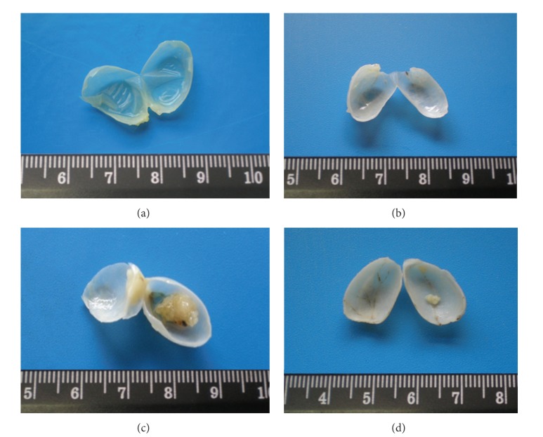
膀胱的宏观组织形态学评估。对照(a)和ω-3-处理的大鼠(b)的所有膀胱均显示正常外观。在BBN组(c)中,65.0%的大鼠表现出旺盛的膀胱癌,几乎占据了膀胱内腔。在这一组中,膀胱表现出显着的血管过度增生,提示肿瘤形成的新血管生成。在预防性ω-3+ BBN组(d)中,发现肿瘤百分率相同,但肿瘤体积明显减少。
N-丁基-N-(4-羟丁基)亚硝胺(BBN)诱导的大鼠膀胱癌实验模型是研究人类癌症发展的合适且经过验证的模型。实际上,由于与人类膀胱癌的组织学相似性,它已成为研究肿瘤病理生理以及评估治疗策略疗效的最常用模型[10-12]。尿路上皮癌变是一个连续而缓慢的过程,它经历了从良性到侵袭性病变的分子和形态变化,包括最初的增生和增生性上皮异常,肿瘤前的变化以及恶性病变(乳头状瘤和癌)[12-14]。因此,假设靶向这些途径的早期治疗可以预防膀胱癌的发展和生长。先前的报告已经证明了预防策略的有益效果,包括我们自己使用抗炎和抗增殖剂进行的研究[15-19]。
多不饱和脂肪酸(PUFA)在体内具有许多生理作用,包括充当细胞能量的来源,蛋白质合成的调节剂,细胞膜结构所需的磷脂和糖脂的基本组成部分,以及调节流动性,渗透性,细胞膜的动力学和动态变化,以及许多激素,细胞间和细胞内的信使及其受体的前体[20,21]。
有多种慢性炎性,氧化性和增殖性疾病,使细胞易于发生肿瘤转化。 ω-3脂肪酸由于其抗氧化和抗炎特性,已被本身作为化学预防剂和/或治疗剂进行了测试,并已与药物和/或放疗结合用于多种癌症[27-30],但关于膀胱癌的推定功效仍有待阐明。因此,本研究的目的是在由亚硝胺诱发的膀胱癌大鼠模型中评估ω-3脂肪酸,特别是二十碳五烯酸(EPA)和二十二碳六烯酸(DHA)的化学预防效力。Omega-3 Fatty Acids Inhibit Tumor Growth in a Rat Model of Bladder Cancer
EXTRACT
Omega-3 (ω-3) fatty acids have been tested on prevention and treatment of several cancer types, but the efficacy on “in vivo” bladder cancer has not been analyzed yet. This study aimed at evaluating the chemopreventive efficacy of eicosapentaenoic acid (EPA) and docosahexaenoic acid (DHA) mixture in an animal model of bladder cancer. Forty-four male Wistar rats were divided into 4 groups during a 20-week protocol: control; carcinogen—N-butyl-N-(4-hydroxybutyl) nitrosamine (BBN); ω-3 (DHA + EPA); and ω-3 + BBN. BBN and ω-3 were given during the initial 8 weeks. At week 20 blood and bladder were collected and checked for the presence of urothelium lesions and tumors, markers of inflammation, proliferation, and redox status. Incidence of bladder carcinoma was, control (0%), ω-3 (0%), BBN (65%), and ω-3 + BBN (62.5%). The ω-3 + BBN group had no infiltrative tumors or carcinoma in situ, and tumor volume was significantly reduced compared to the BBN (0.9 ± 0.1 mm3 versus 112.5 ± 6.4 mm3). Also, it showed a reduced MDA/TAS ratio and BBN-induced serum CRP, TGF-β1, and CD31 were prevented. In conclusion, omega-3 fatty acids inhibit the development of premalignant and malignant lesions in a rat model of bladder cancer, which might be due to anti-inflammatory, antioxidant, anti-proliferative, and anti-angiogenic properties.
The experimental model of rat bladder cancer induced by N-butyl-N-(4-hydroxybutyl) nitrosamine (BBN) is an appropriate and validated model to study human cancer development. In fact, due to the histological similarities with the human bladder cancer, it has been the most used model for the study of tumor pathophysiology, as well as for the evaluation the efficacy of therapeutic strategies [10–12]. The urothelial carcinogenesis is a continuous and slow process that goes through molecular and morphological changes, from benign to aggressive lesions, including initial dysplastic and proliferative epithelial abnormalities, preneoplastic changes, and malignant lesions (papilloma and carcinoma) [12–14]. Thus, an early treatment targeting these pathways could hypothetically prevent bladder cancer development and growth. Previous reports have demonstrated beneficial effects of preventive strategies, including our own studies using anti-inflammatory and anti-proliferative agents [15–19].
Polyunsaturated fatty acids (PUFAs) have many physiological roles in the body, including acting as sources of cellular energy, regulators of protein synthesis, building blocks of phospholipids and glycolipids required for cell membrane structure, and components of membranes that regulate the fluidity, permeability, and dynamics of cell membranes, as well as precursors for many hormones, inter- and intracellular messengers as well as their receptors [20, 21].
There is a wide range of chronic inflammatory, oxidative, and proliferative conditions that make cells susceptible to neoplastic transformation. The omega-3 fatty acids, due to their antioxidant and anti-inflammatory properties have been tested as chemopreventive and/or therapeutic agents, per se and in combination with drugs and/or radiotherapy, in several cancer types [27–30], but the putative efficacy on bladder cancer remains to be elucidated. The purpose of this study was, thus, to evaluate the chemopreventive efficacy of ω-3 fatty acids, in particular eicosapentaenoic acid (EPA) and docosahexaenoic acid (DHA), in a rat model of bladder cancer induced by a nitrosamine.Omega-3 Fatty Acids Inhibit Tumor Growth in a Rat Model of Bladder Cancer
https://www.ncbi.nlm.nih.gov/pmc/articles/PMC3705844/
n-3 PUFA降低人结肠癌细胞中VEGF的表达,从而调节COX-2 / PGE 2诱导的ERK-1和-2以及HIF-1α的诱导途径
n-3多不饱和脂肪酸(PUFA)抑制小鼠乳腺肿瘤中微血管的发育。
人结肠直肠肿瘤产生血管内皮生长因子(VEGF),其表达在肿瘤细胞中被环氧合酶2(COX-2)和PGE 2上调,并与新生血管生成和临床结果直接相关。
这项研究的目的是在体外和体内研究n-3 PUFA调节HT-29人结肠直肠细胞中VEGF表达的能力。HT-29人结肠直肠细胞在减少血清供应是表达VEGF。评估了二十碳五烯酸(EPA)或二十二碳六烯酸(DHA)对VEGF,COX-2,磷酸化细胞外信号调节激酶(ERK)-1和ERK-2和缺氧诱导因子1-α(HIF-1α)的表达和PGE 2的水平的影响。
EPA或DHA喂养的裸鼠中移植的HT-29细胞的肿瘤的生长和VEGF,COX和PGE 2。 EPA和DHA均可降低体外培养的HT-29细胞VEGF和COX-2表达及PGE 2水平。此外,它们抑制ERK-1和-2磷酸化以及HIF-1α蛋白的过度表达,这是PGE 2诱导的信号传导途径中的关键步骤,导致结肠癌细胞中VEGF的表达增加。
EPA在体外总是表现出比DHA更高的功效。两种脂肪酸均降低了通过在裸鼠中接种HT-29细胞获得的肿瘤的生长,微血管形成以及肿瘤中VEGF,COX-2和PGE 2的水平。
数据提供了这些n-3 PUFA能够抑制结肠癌细胞中VEGF表达的证据,并表明涉及的一种可能的机制可能是COX-2 / PGE 2途径的负调控。
提出了其作为抗血管生成化合物在结肠癌治疗中的潜在临床应用。n-3 Polyunsaturated fatty acids (PUFAs) inhibit the development of microvessels in mammary tumors growing in mice.
Institute of General Pathology, 2Institute of Pathology, 3Institute of Pharmacology and 4Institute of Histology, Catholic University, L.go F. Vito, 1, 00168 Rome, Italy
Human colorectal tumors produce vascular endothelial growth factor (VEGF) whose expression is up-regulated in tumor cells by both cyclooxygenase-2 (COX-2) and PGE 2 and directly correlated to neoangiogenesis and clinical outcome.
The goal of this study was to examine the capability of n-3 PUFAs to regulate VEGF expression in HT-29 human colorectal cells in vitro and in vivo .
Constitutive VEGF expression was augmented in cultured HT-29 cells by serum starvation and the effects of eicosapentaenoic (EPA) or docosahexaenoic acid (DHA) on VEGF, COX-2, phosphorylated extracellular signal-regulated kinase (ERK)-1 and -2 and hypoxia-inducible-factor 1-α (HIF-1α) expression and PGE 2 levels were assessed.
Tumor growth, VEGF, COX and PGE 2 analysis were carried out in tumors derived from HT-29 cells transplanted in nude mice fed with either EPA or DHA. Both EPA and DHA reduced VEGF and COX-2 expression and PGE 2 levels in HT-29 cells cultured in vitro . Moreover, they inhibited ERK-1 and -2 phosphorylation and HIF-1α protein over-expression, critical steps in the PGE 2 -induced signaling pathway leading to the augmented expression of VEGF in colon cancer cells.
EPA always showed higher efficacy than DHA in vitro . Both fatty acids decreased the growth of the tumors obtained by inoculating HT-29 cells in nude mice, microvessel formation and the levels of VEGF, COX-2 and PGE 2 in tumors.
The data provide evidence that these n-3 PUFAs are able to inhibit VEGF expression in colon cancer cells and suggest that one possible mechanism involved may be the negative regulation of the COX-2/PGE 2 pathway.
Their potential clinical application as anti-angiogenic compounds in colon cancer therapy is proposed.
n-3 PUFAs reduce VEGF expression in human colon cancer cells modulating the COX-2/PGE 2 induced ERK-1 and -2 and HIF-1α induction pathway | Carcinogenesis | Oxford Academic
https://academic.oup.com/carcin/article/25/12/2303/2475914#authorNotesSectionTitle
Omega-3 Fatty Acids Inhibit Tumor Growth in a Rat Model of ...
https://www.ncbi.nlm.nih.gov/pmc/articles/PMC3705844
Jun 20, 2013 · Omega-3 (ω-3) fatty acids have been tested on prevention and treatment of several cancer types, but the efficacy on “in vivo” bladder cancer has not been analyzed yet.This study aimed at evaluating the chemopreventive efficacy of eicosapentaenoic acid (EPA) and docosahexaenoic acid (DHA) mixture in an animal model of bladder cancer.
Cited by: 14
Publish Year: 2013
Author: Belmiro Parada, Flávio Reis, Raquel Cerejo, Patrícia Garrido, José Sereno, Maria Xavier-Cunha, Paula...
Academic Editor: Achille Cittadini
Omega-3 fatty acid compound found to halt cancerous tumor ...
https://www.naturalnews.com/040931_omega-3_fatty_acids_cancer_tumors.html
Jun 25, 2013 · Omega-3 DHA metabolite stops cancerous metastasis by halting blood supply to tumors The bioactive long-chain omega-3 fat, docosahexaenoic acid (DHA), found in its free form in fatty fish and fish oil supplements, is broken down by the body into a metabolite form known as epoxy docosapentaenoic acid (EDP).
UC Davis Researchers: Key Discovery Involving Omega-3 ...
entomology.ucdavis.edu/News/UC_Davis_Researchers...
Apr 01, 2013 · UC Davis Researchers: Key Discovery Involving Omega-3 Fatty Acids and Cancer ... discovered cytochrome P450 epoxygenase metabolites of omega-3 fatty acid DHA or epoxy docosapentaenoic acids (EDPs) block blood supply to the tumor and ... “The study by Zhang and colleagues has uncovered a previously unrecognized anti-cancer effect of omega-3 ...UC Davis Researchers: Key Discovery Involving Omega-3 Fatty Acids and Cancer - UC Davis Department of Entomology and Nematology
http://entomology.ucdavis.edu/News/UC_Davis_Researchers__Key_Discovery_Involving_Omega-3_Fatty_Acids_and_Cancer/
GPR120 Is an Omega-3 Fatty Acid Receptor Mediating Potent Anti-inflammatory and Insulin-Sensitizing Effects
1
Department of Medicine, Division of Endocrinology and Metabolism, University of California, San Diego, La Jolla, CA 92093, USA
2
Division of Pharmacology, Shiga University of Medical Science, Tsukinowa, Seta, Otsu-city, Shiga, 520-2192 Japan
3
Tethys Bioscience, 3410 Industrial Boulevard, Suite 103, West Sacramento, CA 95691, USA
Summary
Omega-3 fatty acids (ω-3 FAs), DHA and EPA, exert anti-inflammatory effects, but the mechanisms are poorly understood. Here, we show that the G protein-coupled receptor 120 (GPR120) functions as an ω-3 FA receptor/sensor. Stimulation of GPR120 with ω-3 FAs or a chemical agonist causes broad anti-inflammatory effects in monocytic RAW 264.7 cells and in primary intraperitoneal macrophages. All of these effects are abrogated by GPR120 knockdown. Since chronic macrophage-mediated tissue inflammation is a key mechanism for insulin resistance in obesity, we fed obese WT and GPR120 knockout mice a high-fat diet with or without ω-3 FA supplementation. The ω-3 FA treatment inhibited inflammation and enhanced systemic insulin sensitivity in WT mice, but was without effect in GPR120 knockout mice. In conclusion, GPR120 is a functional ω-3 FA receptor/sensor and mediates potent insulin sensitizing and antidiabetic effects in vivo by repressing macrophage-induced tissue inflammation.GPR120 Is an Omega-3 Fatty Acid Receptor Mediating Potent Anti-inflammatory and Insulin-Sensitizing Effects - ScienceDirect
https://www.sciencedirect.com/science/article/pii/S0092867410008883
Akt-mediated regulation of NFκB and the essentialness of ...
https://www.ncbi.nlm.nih.gov/pmc/articles/PMC2767458
Dec 15, 2009 · The serine/threonine kinase Akt (cellular homolog of murine thymoma virus akt8 oncogene), also known as PKB (protein kinase B), is activated by lipid products of phosphatidylinositol 3-kinase (PI3K). Akt phosphorylates numerous protein targets …
Cited by: 281
Publish Year: 2009
Author: Dong Bai, Lynn Ueno, Peter K. Vogt
NF-κB activation by tumour necrosis factor requires the Akt serine–threonine kinase | Nature
Publish Year: 1999
NF-κB activation by tumour necrosis factor requires the Akt serine–threonine kinase
Abstract
Activation of the nuclear transcription factor NF-κB by inflammatory cytokines requires the successive action of NF-κB-inducing kinase (NIK) and an IκB-kinase (IKK) complex composed of IKKα and IKKβ1,2,3,4,5. Here we show that the Akt serine–threonine kinase6 is involved in the activation of NF-κB by tumour necrosis factor (TNF). TNF activates phosphatidylinositol-3-OH kinase (PI(3)K) and its downstream target Akt (protein kinase B). Wortmannin (a PI(3)K inhibitor), dominant-negative PI(3)K or kinase-dead Akt inhibits TNF-mediated NF-κB activation. Constitutively active Akt induces NF-κB activity and this effect is blocked by dominant-negative NIK. Conversely, NIK activates NF-κB and this is blocked by kinase-dead Akt. Thus, both Akt and NIK are necessary for TNF activation of NF-κB. Akt mediates IKKα phosphorylation at threonine 23. Mutation of this amino acid blocks phosphorylation by Akt or TNF and activation of NF-κB. These findings indicate that Akt is part of a signalling pathway that is necessary for inducing key immune and inflammatory responses.
NF-κB activation by tumour necrosis factor requires the Akt serine–threonine kinase | Nature
https://www.nature.com/articles/43466
NF-κB activation by tumour necrosis factor requires the Akt serine–threonine kinase | Nature
https://www.nature.com/articles/43466
Cross-Talk between NFkB and the PI3-Kinase/AKT Pathway Can ...
https://www.ncbi.nlm.nih.gov/pmc/articles/PMC3386924
Jun 29, 2012 · Cross-talk between NFkB pathway and PI3-kinase/AKT pathway has been shown in various cancers , , however, the relationship between these two pathways have not been fully explored in PEL cells. We found that inhibition of NFkB pathway by Bay11-7085 not only inactivates the NFkB pathway, it also inactivates AKT as well as down-stream targets of ...
Cited by: 116
Publish Year: 2012
A
Vitamin C Is a Kinase Inhibitor: Dehydroascorbic Acid ...
https://www.ncbi.nlm.nih.gov/pmc/articles/PMC444845
Vitamin C Is a Kinase Inhibitor: Dehydroascorbic Acid Inhibits IκBα Kinase β Juan M. Cárcamo , 1, 2 Alicia Pedraza , 2 Oriana Bórquez-Ojeda , 2 Bing Zhang , 3 Roberto Sanchez , 3 and David W. Golde 2, *
Cited by: 106
Publish Year: 2004
Author: Juan M. Cárcamo, Alicia Pedraza, Oriana Bórquez-Ojeda, Bing Zhang, Roberto Sanchez, David W. Golde
Vitamin C Is a Kinase Inhibitor: Dehydroascorbic Acid ...
europepmc.org/articles/PMC444845
Vitamin C Is a Kinase Inhibitor: Dehydroascorbic Acid Inhibits IκBα Kinase β Juan M. Cárcamo , 1, 2 Alicia Pedraza , 2 Oriana Bórquez-Ojeda , 2 Bing Zhang , 3 Roberto Sanchez , 3 and David W. Golde 2, *
Vitamin C Is a Kinase Inhibitor: Dehydroascorbic Acid ...
https://www.researchgate.net/publication/8455763_Vitamin_C_Is_a_Kinase_Inhibitor...
Vitamin C Is a Kinase Inhibitor: Dehydroascorbic Acid Inhibits I B Kinase Article in Molecular and Cellular Biology 24(15):6645-52 · September 2004 with 103 Reads How we measure 'reads'
https://content.iospress.com/articles/journal-of-alzheimers-disease/jad170589
Pharmacological Ascorbate-Induced HIF-1α Degradation and VEGF Suppression in Pancreatic Cancer
Justin Wilkes, MD, Juan Du, PhD, Zita Sibenaller, PhD, Adrienne Klinger, Joseph J. Cullen, MD, FACS
University of Iowa, Carver College of Medicine, Iowa City, IAHypoxia-inducible factor-1α (HIF-1α) mediates cellular responses to hypoxia by up-regulating vital homeostatic processes, such as vascular endothelial growth factor (VEGF). In pancreatic adenocarcinoma, HIF-1α strongly affects prognosis and correlates with metastatic disease. Pharmacologic doses of ascorbate (P-AscH-; vitamin C) inhibit pancreatic cancer cell growth by an auto-oxidation reaction resulting in generation of extracellular hydrogen peroxide. We hypothesized that in hypoxic conditions, P-AscH- induces HIF-1α degradation, VEGF suppression, and cytotoxicity.
Pharmacological Ascorbate-Induced HIF-1α Degradation and VEGF Suppression in Pancreatic Cancer - Journal of the American College of Surgeons
https://www.journalacs.org/article/S1072-7515(16)30403-3/abstract
Expression of hypoxia inducible factor 1α and 2α and its ...
https://jbiomedsci.biomedcentral.com/articles/10.1186/s12929-017-0388-y
Oct 30, 2017 · We found an inverse correlation between vitamin C level and HIF-1α but not HIF-2α expression in thyroid lesions. These results agree with our in vitro study showing that vitamin C induced a dose - dependent decrease of HIF-1α but not HIF-2α protein level in …
Cited by: 4
Publish Year: 2017
Author: Paweł Jóźwiak, Piotr Ciesielski, Agnieszka Z
Vitamin C enters mitochondria via facilitative glucose transporter 1 (Glut1) and confers mitochondrial protection against oxidative injury.
Department of Pharmacology, Weill Medical College, Cornell University, New York, New York 10021, USA.
Abstract
Reactive oxygen species (ROS)-induced mitochondrial abnormalities may have important consequences in the pathogenesis of degenerative diseases and cancer. Vitamin C is an important antioxidant known to quench ROS, but its mitochondrial transport and functions are poorly understood. We found that the oxidized form of vitamin C, dehydroascorbic acid (DHA), enters mitochondria via facilitative glucose transporter 1 (Glut1) and accumulates mitochondrially as ascorbic acid (mtAA). The stereo-selective mitochondrial uptake of D-glucose, with its ability to inhibit mitochondrial DHA uptake, indicated the presence of mitochondrial Glut. Computational analysis of N-termini of human Glut isoforms indicated that Glut1 had the highest probability of mitochondrial localization, which was experimentally verified via mitochondrial expression of Glut1-EGFP. In vitro mitochondrial import of Glut1, immunoblot analysis of mitochondrial proteins, and cellular immunolocalization studies indicated that Glut1 localizes to mitochondria. Loading mitochondria with AA quenched mitochondrial ROS and inhibited oxidative mitochondrial DNA damage. mtAA inhibited oxidative stress resulting from rotenone-induced disruption of the mitochondrial respiratory chain and prevented mitochondrial membrane depolarization in response to a protonophore, CCCP. Our results show that analogous to the cellular uptake, vitamin C enters mitochondria in its oxidized form via Glut1 and protects mitochondria from oxidative injury. Since mitochondria contribute significantly to intracellular ROS, protection of the mitochondrial genome and membrane may have pharmacological implications against a variety of ROS-mediated disorders.
https://www.ncbi.nlm.nih.gov/pmc/articles/PMC444845
Hum Mol Genet. 2010 Oct 1;19(19):3721-33. doi: 10.1093/hmg/ddq286. Epub 2010 Jul 16.
Mitochondrial GLUT10 facilitates dehydroascorbic acid import and protects cells against oxidative stress: mechanistic insight into arterial tortuosity syndrome.
Lee YC1, Huang HY, Chang CJ, Cheng CH, Chen YT.
Author information
1
Institute of Biomedical Sciences, Academia Sinica, Taipei 11529, Taiwan, Republic of China.
Abstract
Mutations in glucose transporter 10 (GLUT10) alter angiogenesis and cause arterial tortuosity syndrome (ATS); however, the mechanisms by which these mutations cause disease remain unclear. It has been reported that in most cells, mitochondria are the major source of reactive oxygen species (ROS). Moreover, mitochondria are known to incorporate as well as recycle vitamin C, which plays a critical role in redox homeostasis, although the molecular mechanism(s) underlying mitochondrial vitamin C uptake are poorly understood. We report here that GLUT10 localizes predominantly to the mitochondria of smooth muscle cells and insulin-stimulated adipocytes, where GLUT10 is highly expressed. We further demonstrate that GLUT10 facilitates transport of l-dehydroascorbic acid (DHA), the oxidized form of vitamin C, into mitochondria, and also increases cellular uptake of DHA, which in turn protects cells against oxidative stress. This protection is compromised when GLUT10 expression in mitochondria is inhibited. In addition, we found that aortic smooth muscle cells from GLUT10-mutant mice have higher ROS levels than those from wild-type mice. Our results identify the physiological role of GLUT10 as the mitochondrial DHA transporter, and demonstrate that GLUT10 protects cells from oxidative injury. Furthermore, our findings provide a mechanism to explain the ascorbate in mitochondria and show how loss-of-function GLUT10 mutations may lead to arterial abnormalities in ATS. These results also reinforce the importance of vitamin C and ROS in degenerative diseases.
Mitochondrial GLUT10 facilitates dehydroascorbic acid import and protects cells against oxidative stress: mechanistic insight into arterial tortuos... - PubMed - NCBI
https://www.ncbi.nlm.nih.gov/pubmed/20639396
Vitamin K Inhibits NF-kappaB Activation
https://www.wellnessresources.com/studies/vitamin-k-inhibits-nf-kappab-activation
Furthermore, menaquinone-4, a form of vitamin K2, reduced the activation of nuclear factor κB (NFκB) and inhibited the phosphorylation of IKKα/β after treatment of cells with LPS. These results clearly show that the anti-inflammatory activity of vitamin K is mediated via the inactivation of the NFκB signaling …
Vitamin C blocks TNF-α-induced NF-kB activation and ICAM-1 expression in human neuroblastoma cells
Abstract
Interactions of the cell adhesion molecules are known to play important roles in mediating inflammation. The proinflammatory cytokine, tumor necrosis factor-α (TNF-α), activates the NF-kB signaling pathway, which induces the expression of various genes, such as intercellular adhesion molecule-1 (ICAM-1). In this study, the effect of vitamin C on the ICAM-1 expression induced by TNF-α in a human neuroblastoma cell line, SK-N-SH was investigated. Treatment with vitamin C resulted in the downregulation of the TNF-α-induced surface expression and ICAM-1 mRNA levels in a concentration-dependent manner. Moreover, a gel shift analysis indicated that vitamin C dose-dependently inhibited the NF-kB activation and lkBα degradation induced by TNF-α. Taken together, these results suggest that vitamin C downregulates TNF-α-induced ICAM-1 expression via the inhibition of NF-kB activation.
Vitamin C bloc ks TNF-α-induced NF- kB activation and ICAM-1 expression in human neuroblastoma cellsSpringerLink
https://link.springer.com/article/10.1007/BF02975434
Vitamin C Inhibits NF-κB Activation by TNF Via the Activation of p38 Mitogen-Activated Protein Kinase
Abstract
The transcription factor NF-κB is a central mediator of altered gene expression during inflammation, and is implicated in a number of pathologies, including cancer, atherosclerosis, and viral infection. We report in this study that vitamin C inhibits the activation of NF-κB by multiple stimuli, including IL-1 and TNF in the endothelial cell line ECV304 and in primary HUVECs. The induction of a NF-κB-dependent gene, IL-8, by TNF was also inhibited. The effect requires millimolar concentrations of vitamin C, which occur intracellularly in vivo, particularly during inflammation. Vitamin C was not toxic to cells, did not inhibit another inducible transcription factor, STAT1, and had no effect on the DNA binding of NF-κB. Inhibition by vitamin C was not simply an antioxidant effect, because redox-insensitive pathways to NF-κB were also blocked. Vitamin C was shown to block IL-1- and TNF-mediated degradation and phosphorylation of I-κBα (inhibitory protein that dissociates from NF-κB), due to inhibition of I-κB kinase (IKK) activation. Inhibition of TNF-driven IKK activation was mediated by p38 mitogen-activated protein kinase, because treatment of cells with vitamin C led to a rapid and sustained activation of p38, and the specific p38 inhibitor SB203580 reversed the inhibitory effect of vitamin C on IKK activity, I-κBα phosphorylation, and NF-κB activation. The results identify p38 as an intracellular target for high dose vitamin C.
Vitamin C Inhibits NF-κB Activation by TNF Via the Activation of p38 Mitogen-Activated Protein Kinase | The Journal of Immunology
https://www.jimmunol.org/content/165/12/7180
Quantum chemical study of the autoxidation of ascorbate ...
https://www.researchgate.net/publication/304104302_Quantum_chemical_study_of_the...Ascorbate readily undergoes pH-dependent autoxidation producing hydrogen peroxide (H(2)O(2)). In the presence of catalytic metals this oxidation is accelerated.
Vitamin K Inhibits NF-kappaB Activation
Abstract
Vitamin K is essential for blood coagulation and bone metabolism in mammals. This vitamin functions as a cofactor in the posttranslational synthesis of γ-carboxyglutamic acid (Gla) from glutamic acid residues. However, other functions of vitamin K have been reported recently. We previously found that vitamin K suppresses the inflammatory reaction induced by lipopolysaccharide (LPS) in rats and human macrophage-like THP-1 cells. In this study, we further investigated the mechanism underlying the anti-inflammatory effect of vitamin K by using cultures of LPS-treated human- and mouse-derived cells. All the vitamin K analogues analyzed in our study exhibited varied levels of anti-inflammatory activity. The isoprenyl side chain structures, except geranylgeraniol, of these analogues did not show such activity; warfarin did not interfere with this activity. The results of our study suggest that the 2-methyl-1,4-naphtoquinone ring structure contributes to express the anti-inflammatory activity, which is independent of the Gla formation activity of vitamin K. Furthermore, menaquinone-4, a form of vitamin K2, reduced the activation of nuclear factor κB (NFκB) and inhibited the phosphorylation of IKKα/β after treatment of cells with LPS. These results clearly show that the anti-inflammatory activity of vitamin K is mediated via the inactivation of the NFκB signaling pathway.
Vitamin K Inhibits NF-kappaB Activation
https://www.wellnessresources.com/studies/vitamin-k-inhibits-nf-kappab-activation
The NF-κB Pathway and Cancer Stem Cells
1Department of Pathology and Laboratory Medicine, University of North Carolina, Chapel Hill, NC 27599, USA; ude.cnu.liame@knira
Abstract
The NF-κB transcription factor pathway is a crucial regulator of inflammation and immune responses. Additionally, aberrant NF-κB signaling has been identified in many types of cancer. Downstream of key oncogenic pathways, such as RAS, BCR-ABL, and Her2, NF-κB regulates transcription of target genes that promote cell survival and proliferation, inhibit apoptosis, and mediate invasion and metastasis. The cancer stem cell model posits that a subset of tumor cells (cancer stem cells) drive tumor initiation, exhibit resistance to treatment, and promote recurrence and metastasis. This review examines the evidence for a role for NF-κB signaling in cancer stem cell biology.
Keywords: NF-κB, cancer, cancer stem cells, tumor-initiating cells, self-renewal
The NF-κB Pathway and Cancer Stem Cells
https://www.ncbi.nlm.nih.gov/pmc/articles/PMC4931665/
Ovarian cancer: New drug may prevent recurrence
New research in mice identifies a compound that prevents ovarian cancer recurrence by eradicating the cancer stem-like cells that conventional chemotherapy leaves behind.
New findings could change the face of ovarian cancer treatment.
According to the National Cancer Institute, there were over 22,000 new cases of ovarian cancer in the United States in 2018. More than 14,000 of these resulted in death.
Ovarian cancer is not very common, but its recurrence rate is notoriously high. According to previous estimates, "between 70 and 90 percent of all women with ovarian cancer" will have a recurrence at some point after their diagnosis, depending on how advanced the disease was.
While a person's outlook depends on various factors, just under 50 percent of those who develop ovarian cancer go on to survive for 5 years, according to more recent data.
New research, however, may have found a way to prevent ovarian cancer from coming back. A team of scientists, led by Dr. Ronald Buckanovich, who is also a professor of medicine at the University of Pittsburgh in Pennsylvania, discovered a drug that targets stem-like ovarian cancer cells and stops the cancer from returning.
Dr. Buckanovich and his colleagues published their findings in the journal Cell Reports.
Plucking the root of ovarian cancer
Dr. Buckanovich explains that, even though chemotherapy can destroy up to 99 percent of ovarian cancer cells, the treatment still "misses" stem-like cancer cells.
"You can think of stem-like cells as seeds," explains Dr. Buckanovich, who is also the co-director of the Women's Cancer Research Center at Pittsburgh University. "They put down roots and grow into a plant," he says.
"I especially like the dandelion analogy. When we treat cancer, we're essentially mowing the lawn. But the problem is that dandelions always come back."
The researcher also explains that 11 stem-like cancer cells are enough to form a tumor. So, the researchers set out to find a compound that can eradicate these stem-like cells.
To do so, the scientists carried out a range of chemical experiments. Specifically, they aimed to find a compound that inhibits a pathway known as ALDHA. The cancer cells rely on this pathway to get rid of the toxins that they produce when they replicate quickly.
Study co-author Edward Grimley, Ph.D., who is a Postdoctoral Research Associate in the lab of Dr. Buckanovich, spoke to Medical News Today about the methods the researchers used to find the ALDHA-inhibitor.
Grimley explained that the team screened for a variety of chemical analogs to a "small-molecule known to inhibit the ALDH1A family of enzymes."
"From these experiments, we identified 673A, a potent inhibitor of the ALDH1A family," said Grimley. Then, the researchers showed that this drug efficiently killed cancer stem-like cells in ovarian cancer cell lines.Ovarian cancer: New drug may prevent recurrence
https://www.medicalnewstoday.com/articles/324706.php#1
Surgery for Recurrent Ovarian Cancer Does Not Help Survival - National Cancer Institute
Median overall survival was 50.6 months in the surgery group and 64.7 months in the no-surgery group.
NF-kB promotes ovarian tumorigenesis via classical pathways supporting proliferative cancer cells and alternative pathways supporting ALDH+ cancer stem-like cells
Carrie D House, Elizabeth Jordan, Lidia Hernandez, Michelle Ozaki, Jana M James, Marianne Kim, Michael J Kruhlak, Eric Batchelor, Fathi Elloumi, Margaret C. Cam and Christina M. Annunziata1Women's Malignancies Branch, NCI/NIH
2Medical Oncology Branch, NCI/NIH
3Women's Malignancies Branch, NIH/NCI
4Medical Oncology Branch, National Cancer Institute
5Experimental Immunology Branch, National Cancer Institute
6Laboratory of Pathology, National Cancer Institute
7Developmental Therapeutics Branch, National Institutes of Health
8Neuro-Oncology Branch, National Institutes of Health
Abstract
Understanding the mechanisms supporting tumor-initiating cells (TIC) is vital to combat advanced stage recurrent cancers. Here we show that in advanced ovarian cancers NF-kB signaling via the RelB transcription factor supports TIC populations by directly regulating the cancer stem-like associated enzyme aldehyde dehydrogenase (ALDH). Loss of RelB significantly inhibited spheroid formation, ALDH expression and activity, chemoresistance, and tumorigenesis in subcutaneous and intrabursal mouse xenograft models of human ovarian cancer. RelB also affected expression of the ALDH gene ALDH1A2. Interestingly, classical NF-kB signaling through the RelA transcription factor was equally important for tumorigenesis in the intrabursal model, but had no effect on ALDH. In this case, classical signaling via RelA was essential for proliferating cells, whereas the alternative signaling pathway was not. Our results show how NF-kB sustains diverse cancer phenotypes via distinct classical and alternative signaling pathways, with implications for improved understanding of disease recurrence and therapeutic response.
Received February 3, 2017.
Revision received February 13, 2017.
Accepted October 20, 2017.
Copyright ©2017, American Association for Cancer Research.NF-kB promotes ovarian tumorigenesis via classical pathways supporting proliferative cancer cells and alternative pathways supporting ALDH+ cancer stem-like cells | Cancer Research
https://cancerres.aacrjournals.org/content/early/2017/10/25/0008-5472.CAN-17-0366
Interactions of NF-κB signaling pathway in pancreatic cancer (PC). As indicated in the diagram, both classical and non-classical NF-κB pathways interact with a number of signaling pathways to remain constitutively activated in pancreatic cancer cells and promotes tumorigenesis and metastasis.
NF-κB in pancreatic cancer: Its key role in chemoresistance
Author links open overlay panelQuanxiaoLiab1GangYanga1MengyuFengaSuliZhengaZheCaoaJiangdongQiuaLeiYouaLianfangZhengcYaHuaTaipingZhangadYupeiZhaoa
a
Department of General Surgery, Peking Union Medical College Hospital, Chinese Academy of Medical Sciences and Peking Union Medical College, Beijing, 100730, China
b
Second Affiliated Hospital, Harbin Medical University, Harbin, 150086, China
c
Department of Nuclear Medicine, Peking Union Medical College Hospital, Chinese Academy of Medical Sciences and Peking Union Medical College, Beijing, 100730, China
d
Clinical Immunology Center, Chinese Academy of Medical Sciences, Beijing, 100730, China
Highlights
•
NF-κB activation influences biological behaviors and chemoresistance in PC cells.
•
Activation of NF-κB responding to chemotherapy results in inducible chemoresistance.
•
Side effects need be considered when inhibiting NF-κB in PC adjuvant therapy.
Abstract
Pancreatic cancer is considered a lethal disease with a high mortality and an extremely low five-year survival rate. Chemotherapy plays a pivotal role in pancreatic cancer treatment both in an adjuvant setting after complete resection and in the case of unresectable metastatic disease. However, none of the available combination chemotherapy regimens has resulted in satisfactory survival outcomes. Recent studies have revealed that both constitutive and induced activation of nuclear factor kappa B (NF-κB) in pancreatic cancer cells are closely associated with cell proliferation, invasion, anti-apoptosis, inflammation, angiogenesis and chemotherapeutic resistance. Therefore, NF-κB inhibitors in combination with cytotoxic compounds have been reported as novel agents that improve chemotherapy sensitivity in pancreatic cancer. In this review, we outline recent developments in the understanding of the role of the NF-κB signaling pathway and its associated genes in the progression of pancreatic cancer and highlight some potentially effective strategies for pancreatic cancer treatment.
NF-κB in pancreatic cancer: Its key role in chemoresistance - ScienceDirect
https://www.sciencedirect.com/science/article/abs/pii/S0304383518301381
Cancers (Basel). 2019 Aug; 11(8): 1182.
Published online 2019 Aug 15. doi: 10.3390/cancers11081182
NF-κB Signaling in Ovarian Cancer
Brittney S. Harrington and Christina M. Annunziata*
Women’s Malignancies Branch, National Cancer Institute, Bethesda, MD 20892, USA
Abstract
The NF-κB signaling pathway is a master and commander in ovarian cancer (OC) that promotes chemoresistance, cancer stem cell maintenance, metastasis and immune evasion. Many signaling pathways are dysregulated in OC and can activate NF-κB signaling through canonical or non-canonical pathways which have both overlapping and distinct roles in tumor progression. The activation of canonical NF-κB signaling has been well established for anti-apoptotic and immunomodulatory functions in response to the tumor microenvironment and the non-canonical pathway in cancer stem cell maintenance and tumor re-initiation. NF-κB activity in OC cells helps to create an immune-evasive environment and to attract infiltrating immune cells with tumor-promoting phenotypes, which in turn, drive constitutive NF-κB activation in OC cells to promote cell survival and metastasis. For these reasons, NF-κB is an attractive target in OC, but current strategies are limited and broad inhibition of this major signaling pathway in normal physiological and immunological functions may produce unwanted side effects. There are some promising pre-clinical outcomes from developing research to target and inhibit NF-κB only in the tumor-reinitiating cancer cell population of OC and concurrently activate canonical NF-κB signaling in immune cells to promote anti-tumor immunity.
Keywords: nuclear factor-kappa B, RelA, RelB, stemness, chemoresistance, ovarian cancer
1. Introduction
Ovarian cancer (OC) is the most lethal gynecologic malignancy [1]. The low survival rate of approximately 40% owes to the intra- and inter-tumor heterogeneity, diagnosis at an advanced stage and high rate of recurrence and chemoresistance that classifies this disease [2]. There is increasing evidence that the high rate of disease recurrence after chemotherapy is due to residual populations of cancer stem cells (CSCs) that escaped therapy and re-establish the disease, leading to progression [3]. Populations of CSCs have been identified by certain markers in OC such as CD133, CD44, and ALDH and efforts are focused on strategies to target these populations to prevent recurrent disease [3,4]. The inflammatory but immunosuppressive tumor microenvironment also contributes to OC progression by the production of cytokines that promote survival signaling in OC cells and suppress the infiltration of anti-tumor immune cells [5].
The increased expression of NF-κB signaling molecules had been associated with poor prognosis in many cancers including bladder cancer [6], non-small-cell lung cancer [7] and renal cell carcinoma [8,9]. In OC, the expression levels of NF-κB proteins, p65 and p50, are elevated in malignant and borderline serous ovarian tumors [10,11] and the elevated expression of subunits of the NF-κB pathway, such as the activating IκB kinases (IKKs) β and ε, have been associated with worse overall survival in OC [12,13,14,15]. Constitutive activation of NF-κB was found to be a feature of cell lines derived from granulosa cell tumors of the ovary, thought to be driven by IKKβ; however, the upstream drivers of this pathway were not fully elucidated [16]. In terms of prognostic value, the elevated expression of NF-κB subunits, p65 and p50, has been reported as being greater than 90% of OC cases examined [17,18] and is, therefore, not useful to distinguish patients based on expression of these markers alone. Correlative studies investigating NF-κB pathway proteins and other markers have been reported. The inhibitory proteins that sequester NF-κB in the cytoplasm IκB inversely correlate with p65 expression in OC with poor prognosis, where loss of IκB and increased total p65 expression was associated with worse overall survival [19]. Similarly, the high expression of NF-κB p65 combined with low expression of tumor suppressor PTEN were found to be independent risk factors for chemoresistant ovarian cancer [18]. Another study, focusing on upstream activators of canonical NF-κB signaling in OC, found that high expression of both toll-like receptor 4 (TLR4) and Myd-88 was associated with a poorer outcome and a more aggressive tumor phenotype and was significantly correlated with the expression of NF-κB pathway proteins IKKβ and matrix metalloprotease 9 (MMP9) [17]. The prognostic significance of NF-κB activity in OC is related to its roles in promoting properties of aggressive OC such as chemoresistance [15,20,21,22,23,24] and maintenance of cancer stem cell populations that contribute to recurrence [3,25].
The NF-κB signaling pathway has been implicated in the progression of many cancers, in both blood and solid malignancies [26]. The NF-κB pathway controls multiple cellular processes that can be exploited by cancer cells such as the inhibition of apoptosis in response to chemotherapy, transcription of self-renewal genes in CSCs and the production of anti-tumor cytokines for immune evasion [27]. Due to its role in many normal cellular functions, targeting NF-κB in tumor cells presents a challenge in OC [28]. In this review, we examine the role of the NF-κB signaling pathway in OC chemo-resistance and disease recurrence, inflammation and immunosuppression, and the strategies for targeting this pathway clinically in OC.
2. Canonical and Non-Canonical NF-κB Signaling in OC
The activation of NF-κB signaling in OC has been shown to promote invasiveness, stemness and chemoresistance. NF-κB is a group of transcription factors consisting of five proteins; RelA/p65, c-Rel, RelB, p50 (NF-κB 1) and p52 (NF-κB 2), which are generated by selective degradation in the proteasome of p105 and p100, respectively [28,29]. The composition of the subunits driving NF-κB transcriptional activity directs the signaling towards the canonical or non-canonical pathway. The canonical pathway activates c-Rel-p50 or RelA-p50/p52, while the non-canonical pathway typically activates RelB-p52 [28,29]. Under basal conditions when NF-κB signaling is inactive, the subunits are sequestered in the cytoplasm bound to inhibitory proteins that must be degraded to allow nuclear translocation for transcriptional activity. Inactive c-Rel or RelA/p65 is bound to an inhibitor of κB (IκB) or p105, both of which can be proteasomally degraded after phosphorylation by IκB kinases (IKK) to generate the active RelA/p50 dimer for nuclear translocation and activation of canonical signaling [29,30]. Similarly, RelB is bound to p100 in an inactive state, which can also be phosphorylated by an IKK, but this results in ubiquitination and partial degradation of p100 which generates p52 in the proteasome for nuclear translocation and non-canonical transcriptional activity [29,30].
There are overlapping and distinct activators of the canonical and non-canonical NF-κB signaling pathways and these can differ depending on the cell type [28]. Proinflammatory signaling can activate canonical NF-κB signaling in the innate immune response to pathogens and mitogen-activated protein kinase (MAPK) signaling can enhance cell survival through cellular inhibitors of apoptosis proteins (cIAPs) and canonical NF-κB signaling [29]. Non-canonical NF-κB signaling can be activated in response to adaptive immunity, for B cell activation and expansion and in bone remodeling [31]. Recently, research efforts have focused on delineation of the canonical and non-canonical pathways for their roles in disease progression.
2.1. Canonical NF-κB Signaling in Proliferation and Chemoresistance
The canonical pathway of NF-κB activation has been well established for its anti-apoptotic, pro-angiogenic function in many cancers [32,33]. NF-κB transcription is activated after the degradation of inhibitory IκB proteins by phosphorylation by IKKs, allowing translocation to the nucleus with either RelA/p65 (canonical) or RelB (non-canonical) signaling pathway subunits [25,27,34]. Due to their critical function in the activation of NF-κB signaling, IKKs have been investigated as a target for inhibitors to prevent the nuclear translocation of NF-κB subunits for transcriptional activity. In OC, IKKβ was shown to promote the anchorage-independent growth, proliferation and invasion of OC cells [13]. The pro-angiogenic chemokines IL-8 and VEGF are direct targets of NF-κB and are upregulated by activation of canonical NF-κB signaling through IKKs [13,35,36].
Canonical NF-κB signaling through p65/RelA can be activated in response to inflammatory chemokines and cytokines in the tumor microenvironment. Increasing M2 (pro-tumor) macrophage infiltration and increased p65 phosphorylation and NF-κB activity during tumor progression could be observed intra-vitally in a syngeneic mouse model of OC, using an NF-κB-dependent GFP/luciferase reporter cell line [37]. Inhibition of NF-κB activity in this model was associated with decreased expression of M2 markers and increasing expression of M1 (anti-tumor) markers [37]. This has been confirmed in vitro, where increased expression of p65 is observed in OC cells co-cultured with macrophages, which also showed increased invasiveness and migration compared to OC cells alone [38,39]. Inhibition of NF-κB signaling by inhibitors or RNA interference significantly reduced the invasive phenotype of the co-cultured OC cells [39].
NF-κB activity mediates resistance to stressors of OC cells that occurs during disease progression and metastasis such as oxidative stress and anchorage-independent survival [40]. Inhibition of NF-κB activity in cultured OC cell lines significantly inhibited anchorage-independent growth [40]. Canonical NF-κB signaling regulates the expression of several antioxidant response genes to alleviate elevated cellular reactive oxygen species levels in OC cells [40]. Similarly, numerous studies have shown that the NF-κB pathway is activated in platinum-resistant OC cells to modulate apoptosis and promote cell survival [20,22,41,42,43]. In one example, OC cell lines with resistance to multiple therapies (platinum, paclitaxel and erlotinib) had high NF-κB activity, as measured by p65 phosphorylation, which increased with increasing doses of chemotherapy drugs [21]. Treatment with the NF-κB inhibitor BAY 11-7082 significantly reduced the viability, clonogenicity and anoikis resistance of OC cell lines as a monotherapy in vitro and sensitized cells to multiple therapies [21]. The proposed mechanism of action of BAY 11-7082 appeared to be inhibition of anti-apoptotic genes cIAP1, Bcl-2, and Bcl-xl, direct targets of RelA, and enhanced the expression of p21, a regulator of cell proliferation in the resistant cell lines [21].
DNA damage can also activate NF-κB signaling in a retrograde nuclear to cytoplasmic signaling cascade that is dependent on ataxia telangiectasia-mutated (ATM) [44]. Chemotherapeutic agents that cause genotoxic stress, such as etoposide and doxorubicin, activate NF-κB pro-survival signaling [45,46]. In response to DNA damage, the IKKγ subunit—also known as NF-κB essential modulator (NEMO)—translocates into the nucleus for post-translational modification, phosphorylation by ATM and monoubiquitination for nuclear export [44,47]. In the cytoplasm, the modified NEMO-ATM complex can activate IKKs to phosphorylate and degrade inhibitory IκBα subunits and activate canonical NF-κB survival signaling [44].
Both pro- and anti-apoptotic signaling can be activated downstream of NF-κB in OC cell lines, suggesting a biphasic role for NF-κB in OC progression [24]. In a comparison of chemoresistant cell lines with high NF-κB transcriptional activity with their isogenic, chemosensitive parental cells, it was found that inhibition of the NF-κB pathway using a dominant negative phosphorylation-deficient mutant of IκBα had opposing effects on the chemoresistant and chemosensitive cells of the same origin [24]. The IκBα mutation caused reduced tumor growth of the aggressive, chemoresistant cells in vivo but this same mutation acted to promote tumor growth in the isogenic chemosensitive cells [24]. Apoptosis could be induced in the aggressive chemoresistant cell lines by introduction of this mutation, whereas the chemosensitive cells showed reduced apoptosis [41]. This polar difference in activity and cell viability was related to the ability of NF-κB to differentially regulate mitogen-activated protein kinase (MAPK) phosphorylation to produce pro- or anti- apoptotic signaling in OC cells [24]. This study highlighted the canonical pathway activity and its potential to contribute to chemoresistance and OC cell survival.
Canonical NF-κB signaling may be important in maintaining the proliferative cell populations of tumors, whereas non-canonical NF-κB signaling maintains the CSC populations. In a comparative study of shRNAs targeting RelA or RelB, OC cells grown in tumor-initiating cell (TIC) culture conditions had significantly fewer Ki67 positive, proliferative cell populations and reduced viability with loss of RelA, compared with the loss of RelB [4].
2.2. Non-Canonical NF-κB Signaling and OC Persistence
Activation of the non-canonical NF-κB signaling pathway has been associated with CSC maintenance and self-renewal in OC [4]. A number of genes have been identified as markers of CSCs in OC including CD133, CD117, aldehyde dehydrogenases (ALDH) and CD44 [48,49]. In CD44+ SKOV3 cells there was increased expression of RelA but the expression of non-canonical NF-κB proteins RelB and IKKα was greater, as was the expression of stem cell markers NANOG, SOX2 and OCT4/3 compared to CD44− cells [25]. The CD44+ population showed greater colony-forming capacity in vitro and tumorigenicity in vivo which could be reversed by knocking down IKKα or by expressing a dominant negative mutant in the CD44+ population [25]. In other cancer types, IKKα can be activated and function separately from NF-κB pathway signaling [50,51], but no direct evidence of this has been shown in OC as yet. Thus, the importance of IKKα in this model suggests that the CD44+ CSC population was likely maintained by the non-canonical NF-κB signaling pathway via RelB.
Similar results were reported in the CSC population in other OC cell lines, where both RelA and RelB expression increased in OC cells grown under TIC culture conditions but RelB was more highly expressed in the ALDH+/CD133+ CSC population of OV90 and ACI23 cell lines compared to RelA [4]. Depletion of RelA or RelB in cell line xenografts had differing effects based on the site of tumorigenesis. In subcutaneous and intrabursal xenografts of OC cell lines with inducible knockdown of RelA or RelB with shRNA, loss of either RelA or RelB significantly reduced tumor burden compared to controls [4]. However, intraperitoneal xenografts of shRelA and shRelB cells showed dramatically fewer numbers of tumor cells with shRelB compared to shRelA and control, suggesting there is a greater requirement of non-canonical NF-κB signaling in intraperitoneal, anchorage-independent metastasis in this model [4].
Further evidence of differing roles for canonical and non-canonical NF-κB signaling in OC progression was demonstrated in the context of ECM-interacting protein Transglutaminase (TG2). TG2 can promote OC metastasis in vitro and in vivo and can activate both canonical and non-canonical NF-κB signaling pathways [52,53]. TG2 signaling intracellularly activates canonical NF-κB and a secreted form of TG2 can activate non-canonical NF-κB [52]. In the latter case, RelB activation by secreted TG2 induced CD44 expression, downregulated E-cadherin, and increased invasiveness and peritoneal metastasis in vivo, but interestingly, did not affect cell proliferation in vitro [52]. This is consistent with previous findings about the functions of RelB in OC proliferative and CSC populations [4].
Non-canonical NF-κB signaling is also important for CSC maintenance in endometrioid endometrial cancer (EEC). RelB but not RelA, was significantly upregulated in EEC, and silencing RelB in EEC cell lines affected colony formation, tumor growth in vivo and cell cycle progression [54]. The cells silenced for RelB accumulated in G0/G1, upregulated cell cycle arresting proteins p27 and p21, and activated apoptosis [46]. In comparison, cells with enhanced RelB expression showed advanced cell cycle progression and upregulation of proliferative, apoptosis and cell-cycle-transition-regulating genes such as c-Myc, cyclins D1 and E, and Bcl-2 and Bcl-xl [54].
3. NF-κB Activity in the Inflammatory Ovarian Cancer Microenvironment
The OC microenvironment is unique in that multiple cell types other than the tumor cells are coopted to promote metastasis and inhibit immune activity [5]. There are both inflammatory and immunosuppressive components of the milieu that are controlled by NF-κB activity in the cancer cells, as well as infiltrating immune cells. OC cells secrete immunosuppressive cytokines to promote pro-tumor phenotypes in tumor-associated macrophages (TAMs), which in turn, secrete immunoinhibitory cytokines such as IL-10 and contribute to constitutive NF-κB activation in OC cells by secreting TNFα, promoting survival, CSC expansion and metastasis [59,60].
3.1. Modulation of NF-κB Activity in Infiltrating Immune Cells
TAMs are typically polarized in the pro-tumor ‘M2′ subtype which secrete high levels of IL-10 and TNFα, and decreased IL-12 to promote the immunosuppressive environment and increase invasiveness in cancer cells [61,62]. In a co-culture experiment of mouse bone-marrow-derived macrophages (BMDMs) with mouse ovarian cancer cell line ID-8, the importance of upstream activators of canonical NF-κB signaling was demonstrated by specific knockouts of Myd-88, IL1R, TLR2 and TLR4 in the BMDMs, which caused decreased invasion of ID-8 cells in vitro and decreased tumor growth in vivo [62]. Specific inhibition of the NF-κB classical pathway activating kinase IKKβ in BMDMs caused a shift in the polarization from the pro-tumor M2 subtype to anti-tumor M1 subtype in vitro and in vivo [62]. IKKβ-inhibited BMDMs had decreased production of immunosuppressive cytokines and increased nitric oxide synthesis to promote apoptosis and increased IL-12 production for cytotoxic immune activation [62].
The OC microenvironment contains both pro-inflammatory and immunosuppressive cells so there is a contribution of cytokines by both anti-tumor M1- and pro-tumor M2-polarized macrophages. While M2 macrophages may be recruited locally to the tumor cells to be immunosuppressive, M1 macrophages are also recruited and promote chronic inflammation and high levels of cytokines that are intended to promote cytotoxic T cell recruitment, but these can also act on OC cells to promote survival and metastasis [60,63,64]. For example, treatment with the conditioned medium (CM) of M1-polarized macrophages caused nuclear translocation RelA/p65 and activation of NF-κB transcription, increased invasion and migration in OC cell lines [65]. It was determined that the CM produced by M1-polarized macrophages contained high levels of TNFα and IL-6 and that TNFα activated NF-κB in the OC cells [65].
Dendritic cells are also able to infiltrate the OC microenvironment and can be switched from immunostimulatory to immunosuppressive [66]. A target for cancer immunotherapy is programmed cell death-1 (PD-1), which is a negative regulator of immune cytotoxicity that is expressed on immune cells, particularly T cells [67]. Regulation of immune cytotoxicity through PD-1 is directed by interaction with its ligand (PD-L1), which is expressed on normal cells but has also been detected on cancer cells as a strategy to avoid immune cytotoxicity [67]. DCs from OC tumors and ascites that expressed PD-1 had decreased anti-tumor activity due to inhibition of canonical, immunogenic NF-κB activity [68]. Blockade of PD-1 on the DCs restored their pro-inflammatory functions by activation of canonical NF-κB with degradation of IκBα, allowing the nuclear translocation of RelA/p65 [68]. In the PD-1-blocked DCs, the upregulation of co-stimulatory molecules and secretion of immunogenic and proinflammatory cytokines TNFα, Il-6 and G-CSF was found to be dependent on NF-κB [68]. Unfortunately, the co-activation of NF-κB activation in OC cells in response to these cytokines can be utilized to further promote the immunosuppressive pro-tumor environment.
3.2. NF-κB Activation in Ovarian Cancer Cells to Alter the Microenvironment
OC cells contribute to the maintenance of the tumor microenvironment by secreting immunosuppessive cytokines to prevent migration of anti-tumor immune cells and to polarize macrophages to a pro-tumor M2 subtype [61,69]. This phenomenon triggers the expression of multiple cytokines including TNFα from tumor-infiltrating macrophages exposed to OC cells [61]. Accordingly, constitutive NF-κB signaling is activated in OC cells in response to co-culture with M2 macrophages and in response to high TNFα [38,69]. In addition, constitutively activated NF-κB signaling in OC cells also leads to increased immunosuppressive cytokines to further promote tumor growth [70]. The activation of canonical NF-κB signaling in OC cells is a major component of the immunosuppressive cytokine secretion, and inhibition of the pathway in OC cells caused the immune cell infiltrates to switch to an anti-tumor phenotype in vitro and in vivo [70,71,72].
Persistent inflammation has been associated with ovarian cancer development, in women with pelvic inflammatory disease or in response to pathogenic infections of Chlamydia trachomatis or Mycoplasma genitalium [73]. Recognition of pathogen-associated molecular patterns (PAMPs) by cell receptors such as TLRs initiates the innate immune response by activating NF-κB, which stimulates the production of pro-inflammatory cytokines including TNFα, IL-6, IL-8, and MCP-1 [74]. Persistent, chronic inflammation following infection and constitutive NF-κB activity may contribute to ovarian carcinogenesis by producing pro-inflammatory and pro-angiogenic cytokines and through anti-apoptotic signaling [73].
Another way OC cells use NF-κB signaling for immune evasion is through PD-L1 upregulation [75]. In response to chemotherapy, the expression of p65/RelA and PD-L1 increased in OC cell lines in vitro and in vivo [75]. Silencing p65/RelA prevented PD-L1 upregulation, confirming its dependence on NF-κB activity [75]. Thus, the OC microenvironment activates NF-κB signaling in a variety of cell types to promote tumor cell survival, immune evasion and metastasis.
Figure 1
Strategies to target NF-κB in ovarian cancer. NF-κB signaling in cancer cells can be activated by interacting proteins in the tumor microenvironment, such as lysophosphatidic acid (LPA) signaling through CD44 to promote NF-κB activity and cytokine secretion. NF-κB signaling can also be activated by TNFα binding to cell surface receptors Myd-88 and TLR4, or through TNFα independent receptors such as TG2. NF-κB activity promotes cytokine secretion to dampen immune responses and switch macrophages from inflammatory M1 phenotypes to M2 immunoinhibitory phenotypes. Selective activation of NF-κB signaling in dendritic cells and macrophages with toll-like receptor (TLR) agonists or blockade of PD-1 could promote anti-tumor immunity and recruitment of additional anti-tumor immune cells. Alternatively, selective inhibition of NF-κB in tumor cells by IκB kinase (IKK) inhibitors could attenuate pro-tumor survival signaling in the tumor cells without disrupting inflammatory NF-κB activity in immune cells.NF-κB Signaling in Ovarian Cancer
5. Conclusions
The NF-κB signaling pathway has been implicated in OC tumorigenesis through its roles in mediating chemoresistance, maintenance of CSC populations, metastasis and immune evasion. The NF-κB signaling proteins and IKKs are overexpressed in OC tumors and correlate with poorer patient outcomes. Both canonical and non-canonical NF-κB signaling contribute to OC progression with overlapping and distinct roles in promoting chemoresistance and CSC maintenance. The inflammatory tumor microenvironment is also heavily influenced by NF-κB signaling, from the inflammatory cytokines that activate NF-κB signaling in OC cells and the immunosuppressive phenotype of TAMs and other immune cells that have diminished NF-κB activity. Targeting NF-κB in OC to eliminate the CSC population is desirable, but current strategies are limited to inhibition of IKKs. More research is needed to dissect the pathways and identify strategies to modulate NF-κB to reduce metastasis and tumor recurrence. There is a need to strike a balance between the inhibition of the non-canonical NF-κB in OC cells which enables long-term resistance and tumor re-initiation from CSC and the activation of canonical NF-κB in immune cells to generate an effective anti-tumor immune response.
https://www.ncbi.nlm.nih.gov/pmc/articles/PMC6721592/
BMC Complement Altern Med. 2007; 7: 44.
Published online 2007 Dec 20. doi: 10.1186/1472-6882-7-44
Ginger inhibits cell growth and modulates angiogenic factors in ovarian cancer cells
Jennifer Rhode,#1 Sarah Fogoros,#2 Suzanna Zick,3 Heather Wahl,4 Kent A Griffith,5 Jennifer Huang,4 and J Rebecca Liucorresponding author4
188th Medical Group, Wright-Patterson AFB, Ohio, USA
2National Institutes of Health, National Human Genome Research Institute, Cancer Genetics Branch, USA
3Department of Family Medicine, University of Michigan, Ann Arbor, MI, USA
4Department of Obstetrics and Gynecology, Division of Gynecologic Oncology, University of Michigan, Ann Arbor, MI, USA
5Department of Biostatistics, University of Michigan, Ann Arbor, MI, USA
corresponding authorCorresponding author.
#Contributed equally.
Jennifer Rhode: moc.loa@edohRMnJ; Sarah Fogoros: vog.hin.liam@sorogof; Suzanna Zick: ude.hcimu.dem@kcizs; Heather Wahl: ude.hcimu@lhawh; Kent A Griffith: ude.hcimu@gtnek; Jennifer Huang: ude.hcimu@hynej; J Rebecca Liu: ude.hcimu@uilr
This article has been cited by other articles in PMC.
Go to:
Abstract
Background
Ginger (Zingiber officinale Rosc) is a natural dietary component with antioxidant and anticarcinogenic properties. The ginger component [6]-gingerol has been shown to exert anti-inflammatory effects through mediation of NF-κB. NF-κB can be constitutively activated in epithelial ovarian cancer cells and may contribute towards increased transcription and translation of angiogenic factors. In the present study, we investigated the effect of ginger on tumor cell growth and modulation of angiogenic factors in ovarian cancer cells in vitro.
Methods
The effect of ginger and the major ginger components on cell growth was determined in a panel of epithelial ovarian cancer cell lines. Activation of NF-κB and and production of VEGF and IL-8 was determined in the presence or absence of ginger.
Results
Ginger treatment of cultured ovarian cancer cells induced profound growth inhibition in all cell lines tested. We found that in vitro, 6-shogaol is the most active of the individual ginger components tested. Ginger treatment resulted in inhibition of NF-kB activation as well as diminished secretion of VEGF and IL-8.
Conclusion
Ginger inhibits growth and modulates secretion of angiogenic factors in ovarian cancer cells. The use of dietary agents such as ginger may have potential in the treatment and prevention of ovarian cancer.
Results
Ginger inhibits growth in ovarian cancer cells as compared to non-transformed ovarian epithelial cells
Continuous exposure to ginger extract resulted in a marked reduction in cell growth after 1–5 days of exposure in A2780 ovarian cancer cells (Figure (Figure1A,1A, p < .0001 at all doses and time points). We tested additional ovarian cancer cell lines to determine if this was an effect unique to the A2780 ovarian cancer cells. Ginger treatment resulted in similar effects in all cell lines tested, including the chemoresistant cell lines SKOV3 and ES-2[39] (Figure 1B,C, p < .05 for all doses and time points). Untransformed human ovarian surface epithelial cells (HOSE) were minimally affected by ginger extract exposure at days 1 and 3, and showed some inhibition in growth by day 5 (Figure (Figure1D,1D, p > .05 for days 1 and 3, p < .05 for day 5). To confirm that ginger treatment inhibited cell growth, treated cells were analyzed by trypan blue exclusion as well. As expected, ginger treatment resulted in a profound inhibition of cell proliferation and growth at doses of 50 μg/ul and higher (Figure (Figure22).
Continuous ginger exposure inhibits growth in ovarian cancer cells in vitro. A, B, C: The effect of ginger on growth of A2780, SKOV3 and ES2 ovarian cancer cell lines was assessed by using sulforhodamine B assays. Cells were incubated continuously with media containing ginger at the indicated concentrations and growth was assayed at Days 1, 3 and 5 of exposure. Ginger treated cells displayed significant growth inhibition as compared to control treated cells (p < .05 for all cell lines, all ginger concentrations). D: Human ovarian surface epithelial cells were treated with the indicated concentrations of ginger with minimal effect seen following days 1–3 of culture (p > .05). HOSE demonstrated some inhibition of growth by day 5 of treatment (p < .05). Data are presented as means ± S.D, and are representative of at least 3 independent experiments.
Figure 2
Continuous ginger exposure inhibits growth in ovarian cancer cells in vitro. A, B, C: The effect of ginger on growth of A2780, SKOV3 and ES2 ovarian cancer cell lines was assessed using the trypan blue exclusion assay. Cells were incubated continuously with media containing ginger at the indicated concentrations and viable cells were counted on days Days 1, 3 and 5 of exposure. Ginger treated cells displayed significant growth inhibition as compared to control treated cells.
To determine whether lower doses of ginger could also inhibit cell growth, an extended range of concentrations was tested. In the A2780 and ES-2 cell lines, ginger concentrations of less than 50 μg/ml did not significantly impact cell growth, whereas in the SKOV3 cell line, some inhibition of cell growth was seen with ginger concentrations as low as 30 μg/ml (Figure (Figure33 and data not shown).
6-Shogaol is the most active of the individual ginger components tested in ovarian cancer cells
Previous investigators have shown bio-activity of various individual ginger components in several tumor types [30,40-43]. To determine the relative bio-activity in ovarian cancer, A2780 ovarian cancer cells were treated with 6-, 8- and 10-gingerol as well as 6-shogaol. In contrast to other published findings, we found that 6-, 8- and 10-gingerol had no effect on the growth or viability of ovarian cancer cells (p > .05 at all time points). Treating cells with whole ginger extract or 6-shogaol resulted in profound growth inhibition (Figure (Figure4A,4A, p < .05 at all time points for both ginger and 6-shogaol treated cells). Morphologically, cells treated with ginger appeared markedly growth inhibited, similar to cisplatin treated cells (Figure (Figure5).5). Cells cultured with vehicle control (DMSO) continued to proliferate.
Figure 5
Morphologic appearance of ginger treated ovarian cancer cells. A2780 ovarian cancer cells were incubated with DMSO solvent, ginger (75 μg/ml, replenished on day 3), or Cisplatin (2.5 μg/ml). Cells were examined by light microscopy at 1, 3, and 5 days of treatment.
We next determined if continuous exposure to individual ginger components was necessary to cause the growth inhibitory effect seen in ovarian cancer cells. Similar to the use of whole ginger root extract, continuous exposure to 6-shogaol was necessary to cause the growth inhibitory effect seen in ovarian cancer cells. We treated cells with ginger and individual ginger components for 24 hours, after which the cells were washed and media was changed. Once ginger or 6-shogaol was removed from the media, cell growth resumed (Figure (Figure4B4B).
Ginger inhibits NF-κB in ovarian cancer cells
Because we found that ginger markedly suppressed ovarian cancer cell proliferation in vitro, and several genes that regulate proliferation are regulated by NF-κB, we hypothesized that ginger may mediate its anti-neoplastic activity in ovarian cancer cells though modulation of this pathway. Constitutive activation of NF-κB has been described in many tumor types including ovarian cancer[9], suggesting that targeting NF-κB may have an anti-neoplastic effect in this tumor type. Natural products such as ginger, or ginger components such as zerumbone can inhibit NF-κB in other cell types [16,44,45]. We chose two chemoresistant ovarian cancer cell lines (CaOV3 and SKOV3) to evaluate the effect of ginger treatment on activation of NF-κB. As shown in Figure Figure6,6, treatment with ginger extract resulted in a significant inhibition of NF-κB activation in CaOV3 and SKOV3 cell lines
Figure 6
Ginger inhibits NF-kB activation in ovarian cancer cells. Representative ovarian cancer cell lines with high endogenous NF-kB activation, (A) CaOV3 and (B) SKOV3 were transfected with an NF-κB-dependent reporter plasmid (pBVIx-Luc). Cells were treated with DMSO (vehicle control) or ginger (75 μg/ml). NF-κB activation was determined by measuring relative luciferase activity 48 hours after treatment. Luciferase activity is reported as arbitrary relative light units (mean +/- S.D.) Ginger treatment resulted in inhibition of NF-κB activation (p < .05 for both cell lines). Representative data is shown.
Ginger Inhibits IL-8 and VEGF Secretion in Ovarian Cancer Cells
IL-8 can function as a paracrine and/or autocrine growth factor in some tumor types, and the secretion of IL-8 protein from tumor cells themselves is thought to be crucial for these effects[46,47]. In ovarian cancer patients, elevated IL-8 expression has been found in ascites as well as in serum[33]. Furthermore, IL-8 has been shown to stimulate proliferative growth in ovarian cancer cells in vitro[35]. Because IL-8 secretion is thought to be regulated in part by NF-κB, and ginger can clearly inhibit NF-κB in ovarian cancer cells, we hypothesized that ginger could also inhibit IL-8 secretion. Using a representative panel of ovarian cancer cell lines, we found that A2780 and CaOV3 cells produced negligible amounts of IL-8 (<0.05 pg/ml), whereas the cell lines ES-2 and SKOV3 had high constitutive expression of IL-8 (Figure (Figure7A).7A). Treatment with ginger resulted in significant inhibition of IL-8 production in the ES-2 and SKOV3 cell lines (p < .05 for both cell lines).
Figure 7
Ginger inhibits VEGF and IL-8 in ovarian cancer cells. Ovarian cancer cells were cultured with DMSO (vehicle control) or Ginger (75 μg/ml) for 48 hours. Production of the angiogenic factor s IL-8 (A) and VEGF (B) were assayed using ELISA assays. (A.) Only ES-2 and SKOV3 cells expressed high IL-8 levels at Baseline, and ginger treatment resulted in a significant decrease in IL-8 production (p < .05 for both cell lines). (B.) VEGF production was reduced in all cell lines following ginger treatment (p = .19, .18, .007, and .07 for A2780, CaOV3, ES-2 and SKOV3 cells respectively).
VEGF, the most important inducer of angiogenesis, is also under transcriptional control of NF-κB[9]. Serum VEGF levels as well as tumor expression of VEGF are associated with poor prognosis in ovarian cancer patients [31], and inhibition of VEGF function using Avastin™ has shown promise in the treatment of ovarian cancer patients[48]. Because ginger treatment resulted in inhibition of NF-κB, we next sought to determine whether ginger could similarly inhibit VEGF in ovarian cancer cells. In all cell lines tested, there was high endogenous production of VEGF, and ginger treatment resulted in inhibition of VEGF secretion. Inhibition of VEGF secretion was most evident in the ES-2 cell line (p = .007), as compared to the other cell lines tested (p = .19, .18, and .07 for A2780, CaOV3, and SKOV3 respectively, Figure Figure7B7B).
Discussion
The analysis of epidemiologic data and disparities in global incidence of ovarian cancer may provide clues to uncover environmental and biologic factors that contribute towards the development of ovarian cancer. Dietary prevalence of foods such as ginger, garlic, soy, curcumin, chilies and green tea are thought to contribute to the decreased incidence of colon, gastrointestinal, prostate, breast and other cancers in South East Asian countries [49]. Accumulating evidence suggests that many dietary factors may be used alone or in combination with traditional chemotherapeutic agents to prevent or treat cancer. The potential advantage of many natural or dietary compounds seems to focus on their potent anticancer activity combined with low toxicity and very few adverse side effects.
Epithelial ovarian carcinoma is the leading cause of death among patients with gynecologic cancers. Despite multiple modalities of treatment including surgery and chemotherapy, ovarian cancer patients continue to have one of the lowest 5-year survival rates [1]. The significant morbidity and limited success of surgery and chemotherapy for ovarian cancer has led to searches for alternative therapies. Recently, ginger root and its main poly-phenolic constituents (gingerols and zerumbone) been shown to exhibit anti-inflammatory [16-19], and anti-neoplastic activity [20-24] in several cell types through inhibition of the transcription factor NF-κB [25-28]. NF-κB plays an important role in tumorigenesis, given its ability to control the expression and function of numerous genes involved in cell proliferation, sustained angiogenesis, and evasion of apoptosis. Different tumor types, including ovarian cancer, have been shown to express high constitutive NF-κB activity[9]. In this study we show that ginger blocks NF-κB activation in ovarian cancer cells, resulting in inhibition of NF-κB regulated gene products involved in cellular proliferation and angiogenesis.
Many of the pathways that mediate adaptive survival strategies in cancer cells are under the transcriptional control of NF-κB [9]. We have shown here that in ovarian cancer cells, NF-κB is constitutively activated, and blocking NF-κB activation with ginger results in suppressed production of NF κB regulated angiogenic factors and selectively inhibits ovarian cancer cell growth. We have found that ginger selectively inhibits ovarian cancer cell growth, as compared to non-transformed ovarian epithelial cells. Previous reports indicate that the ginger component 6-shogaol induces cell death in chemoresistant hepatoma cells [50], yet inhibits cell death in non-neoplastic spinal cord cells [51], suggesting that ginger and ginger components' effects are cell type specific. The apparent contradictory findings may be due to a differential effect of ginger on transformed cells (i.e. cancer cells) vs. untransformed cells. Phytochemicals such as ginger, generally have multiple molecular targets. This pleiotropism may constitute an advantage in the treatment of ovarian cancer, where multiple factors contribute towards the carcinogenic process.
Conclusion
The results of this study indicate that ginger may exhibit anti-neoplastic effects through the inhibition of NF-κB. Further studies utilizing ginger in an in vivo model of ovarian cancer will provide a platform for the development of ginger as a therapeutic tool in this disease.Ginger inhibits cell growth and modulates angiogenic factors in ovarian cancer cells
https://www.ncbi.nlm.nih.gov/pmc/articles/PMC2241638/
Zingiber officinale attenuates retinal microvascular changes in diabetic rats via anti-inflammatory and antiangiogenic mechanisms. - PubMed - NCBI
https://www.ncbi.nlm.nih.gov/pubmed/27293376
The NFκB luciferase mouse: a new tool for real time ...
https://www.ncbi.nlm.nih.gov/pmc/articles/PMC1773458
The NFκB luciferase mouse: a new tool for real time measurement of NFκB activation in the whole animal D A Mann Liver Research Group, University of Southampton, Southampton, UK; Email: ku.ca.notos@2mad
Gut. 2002 Dec; 51(6): 769–770.
The NFκB luciferase mouse: a new tool for real time measurement of NFκB activation in the whole animal
D A Mann
Liver Research Group, University of Southampton, Southampton, UK; ku.ca.notos@2mad
Of the many different transcription factors found in mammalian cells by far the best known among scientists and clinicians alike is nuclear factor κB (NFκB). In addition to its pivotal role as a regulator of innate and adaptive immune responses, NFκB has also been linked as a contributory factor in many human disorders.1 NFκB stimulates transcription of over 150 different genes encoding cytokines, adhesion molecules, immunoreceptors, acute phase proteins, enzymes, and regulators of the cell cycle and apoptosis.2 Transcription of these genes is stimulated in response to binding of NFκB to a specific 10 base pair DNA sequence (GGGRNNYYCC, where R corresponds to a purine, Y represents a pyrimidine, and N can be any base) located in either the enhancer or upstream promoter regions of a gene. Binding of NFκB to these sequences is tightly regulated by the inhibitory function of the IκB-α protein which forms a complex with NFκB that masks functional domains of the transcription factor required for its nuclear localisation and DNA binding activities. The mechanism by which IκB-α bound NFκB is activated is now well understood (fig 1 ▶).3 Following stimulation of the cell by an extracellular signal, such as the interaction of tumour necrosis factor α with its receptor, the recently discovered IκB kinase (IKK) complex is activated. The IKK complex then catalyses phosphorylation of IκB-α at two N terminal serine residues (ser 32 and 36) which then renders IκB-α a substrate for ubiquitination and finally for degradation by the 26S proteasome. These events are rapid, and within minutes of the initial stimulation event result in the nuclear translocation of active NFκB. A remarkable number of different inducers of the NFκB pathway have been described, including a plethora of cytokines and a surprising variety of different viral and bacterial gene products.2 Given the ability of NFκB to be activated in response to this diverse range of extracellular stimuli and its subsequent transcriptional activation of a vast number of target genes, it is not surprising that the transcription factor has been implicated in so many disease processes. Gastrointestinal diseases in which NFκB has been linked as a contributor to pathogenesis include fibrogenesis, inflammatory bowel disease, acute pancreatitis, responses to infections by bacteria and viruses, and last but not least cancer.4
Figure 1
Events leading to activation of nuclear factor κB (NFκB) dependent gene transcription. Following the interaction of a cell surface receptor with its ligand (for example, tumour necrosis factor α (TNF-α)), the IκB kinase (IKK) complex is activated resulting in phosphorylation (P), ubiquitination (Ub), and proteasome mediated degradation of IκB-α. Free NFκB (p65:p50) dimers then translocate to the nucleus where additional phosphorylation events on p65 and recruitment of the coactivator and histone acetyltransferase CBP/p300 leads to transcriptional activation of up to 150 different genes. INOS, inducible nitric oxide synthase; COX2, cyclooxygenase 2.
Carlsen H, Moskaug JØ, Fromm SH, et al. In vivo imaging of NF-kappa B activity. J Immunol 2002;168:1441–6.
A wide range of human disorders involves inappropriate regulation of NF-kappaB, including cancers and numerous inflammatory conditions. Toward our goal to define mechanisms through which NF-kappaB leads to the development of disease, we have developed transgenic mice that express luciferase under the control of NF-kappaB, enabling real-time in vivo imaging of NF-kappaB activity in intact animals. We show that in the absence of extrinsic stimulation, strong luminescence is evident in lymph nodes in the neck region, thymus, and Peyer’s patches. Treating mice with TNF-alpha, IL-1alpha, or LPS increased the luminescence in a tissue-specific manner, with the strongest activity observed in skin, lungs, spleen, Peyer’s patches, and the wall of the small intestine. Liver, kidney, heart, muscle, and adipose tissue displayed less intense activities. Also, exposure of skin to a low dose of UV radiation increased luminescence in the exposed areas. Furthermore, induction of chronic inflammation resembling rheumatoid arthritis produced strong NF-kappaB activity in the affected joints, as revealed by in vivo imaging. Thus, we have developed a versatile model for monitoring NF-kappaB activation in vivo.
To date, most of what has been inferred about the potential role played by NFκB in gastrointestinal disease has been derived from a combination of in vitro and ex vivo studies measuring changes in NFκB nuclear localisation or DNA binding activity. The problem with these studies is that they are based on the often incorrect assumption that the transcriptional activity (or trans-activation) of NFκB can be directly correlated with its degree of nuclear location and DNA binding. Similarly, the suggestion that many commonly used anti-inflammatory drugs including aspirin, sulphasalazine, and glucocorticoids influence the activation of NFκB is largely based on this assumption. In fact, the degree of trans-activation by NFκB is also influenced by additional regulatory mechanisms including post-translational modifications (for example, phosphorylation) of NFκB and its interaction with transcriptional coactivators such as CBP/p300.5 The paper by Carlsen et al describes the production of a new experimental tool that provides a significant advance in our ability to quantify changes in NFκB dependent transcription in real time, in a living animal and in a relatively non-invasive manner. What Carlson et al have produced is a transgenic mouse that carries in its genome a firefly luciferase gene under the control of three tandem NFκB DNA binding sites. Consequently, this mouse will express luciferase in any cell in which active NFκB is present; moreover, the level of luciferase expressed is proportional to the level of NFκB trans-activation in a cell or tissue. Measurement of luciferase expression is achieved by intravenous injection of the animal with a suitable substrate such as d-luciferin which in the presence of the enzyme leads to a bioluminescent reaction that can be detected in the whole animal using an image intensifier coupled to a CCD camera. As firefly luciferase has only a short biological half-life (two hours), the system is capable of measuring temporal fluctuations in NFκB activity which is a tremendous advance over other systems such as DNA binding assays that only provide a static snapshot of NFκB activity. But perhaps the most impressive advantage is that the system is sufficiently sensitive to image and quantify NFκB activity within the internal organs (including the small intestine and the liver) of the intact animal.
How does the availability of the NFκB luciferase mouse impact on gastroenterology? The answer is that for the first time we can now properly monitor the influence of exogenous agents and pathological conditions on NFκB activity in the gut and liver. This includes the potential to screen for new drugs that target aberrant NFκB activity. It should also be possible to accurately determine the effective dose and administration regimen for a given anti-NFκB drug in an organ specific manner. Rather than simply determining the delivery and uptake of these drugs into the gut and liver, the NFκB luciferase mouse will now tell us the biological efficacy of the drug within the tissue and over what timeframe the drug can be expected to remain effective. As well as impacting on clinical research the NFκB reporter mouse has tremendous potential in basic research, especially when combined with experimental in vivo models of disease. One of the major problems associated with studying NFκB is its amazing sensitivity to changes in the cellular environment; the very process of isolating tissues and organs from an animal may itself alter NFκB activity. To date it has therefore been virtually impossible to determine changes in the transcriptional activity of NFκB during a disease process. Carrying out in vivo models of gastrointestinal disease in the NFκB luciferase mouse will enable us to accurately quantify the degree to which NFκB dependent transcription is increased throughout the entire course of a disease. Such studies will significantly improve our understanding of the role played by NFκB in gastrointestinal diseases.
The NFκB luciferase mouse: a new tool for real time measurement of NFκB activation in the whole animal
https://www.ncbi.nlm.nih.gov/pmc/articles/PMC1773458/
NFkB Luciferase Reporter HeLa Stable Cell Line
Description:
NFkB Responsive Luciferase Reporter Hela Stable Cell Line is derived from human cervical cancer, and stably express firefly luciferase reporter gene under the control of the NFkB response element. This cell line is an ideal cellular model for monitoring the activation of NFkB Pathway triggered by stimuli treatment, enforced gene expression and gene knockdown.
Principle
NFkB plays an important role in controlling many biological processes including immune and inflammatory responses, developmental processes, cellular growth, and apoptosis. In response to the various stimuli, such as stress, cytokines, free radicals, ultraviolet irradiation, and bacterial or viral antigens, NFkB is activated and translocate from cytoplasm to nucleus, where NFkB binds to its response element on the promoter region and regulates a wide spectrum of gene expression. Dysfunction of NFkB activity is associated with cancer, inflammatory and autoimmune disease, and viral infection. Monitoring the NFkB activity is essential to unveil the mechanism of these diseases and conduct drug discovery. Signosis has established NFkB luciferase reporter Hela stable cell line that has been stably transfected with pTA-NFkB-luciferase reporter vector, which contains 4 repeats of NFkB binding sites, a minimal promoter upstream of the firefly luciferase coding region. Therefore, the cell line can be used as a reporter system for monitoring the activation of NFkB triggered by stimuli treatment, enforced gene expression and gene knockdown. The stable cell line can be used for studying NFkB signaling pathways activated by different cytokines, such as TNFa, and many other stimuli.
The cell line was established by transfection using a pTA-NFkB-luciferase reporter vector, which contains 4 repeats of NFkB binding sites, a minimal promoter upstream of the firefly luciferase coding region, along with hygromycin expression vector followed by hygromycin selection. The hygromycin resistant clones were subsequently screened for TNFa-induced (or IL-1) luciferase activity.
Analysis of TNFa/NFkB Pathway Reporter Cell Line in response to TNFa treatment. The cells were seeded on a 96-well plate for overnight with DMEM including 10% FBS. The cell then were treated with 20ng/ml TNFa in DMEM + 0.1% FBS for 6 hours.
Principle behind TF luciferase reporter. TF luciferase reporter stable cell line utilizes artificial promoter constructs to drive luciferase expression. The promoter region can consists of multiple repeats of a cis-element TF binding site, a DNA fragment from the promoter region of a known TF downstream gene, or a DNA fragment containing putative/known TF binding sites. There are several ways that a TF can be activated, such as through extracellular stimuli or through intracellular signaling pathways. Once activated, the TF translocates to the nucleus and often interacts with relevant co-factors to drive gene expression. Once luciferase is expressed, it can generate light in an enzymatic assay and the amount of light measured is positively correlated with the level of TF activation.
Signosis
https://www.signosisinc.com/product/nfkb-luciferase-reporter-hela-stable-cell-line-sl-0001-np
Published: 14 September 2017
Endogenous ω-3 Fatty Acid Production by fat-1 Transgene and Topically Applied Docosahexaenoic Acid Protect against UVB-induced Mouse Skin Carcinogenesis
Hye-Won Yum, Jin Park, Hyun-Jung Park, Jun Wan Shin, Yong-Yeon Cho, Su-Jung Kim, Jing X. Kang & Young-Joon Surh
Scientific Reports volume 7, Article number: 11658 (2017) Cite this article
Abstract
The present study was intended to explore the effects of endogenously produced ω-3 polyunsaturated fatty acids (PUFAs) on ultraviolet B (UVB)-induced skin inflammation and photocarcinogenesis using hairless fat-1 transgenic mice harboring ω-3 desaturase gene capable of converting ω-6 to ω-3 PUFAs. Upon exposure to UVB irradiation, fat-1 transgenic mice exhibited a significantly reduced epidermal hyperplasia, oxidative skin damage, and photocarcinogenesis as compared to wild type mice. The transcription factor, Nrf2 is a master regulator of anti-inflammatory and antioxidant gene expression. While the protein expression of Nrf2 was markedly enhanced, the level of its mRNA transcript was barely changed in the fat-1 transgenic mouse skin. Topical application of docosahexaenoic acid (DHA), a representative ω-3 PUFA, in wild type hairless mice induced expression of the Nrf2 target protein, heme oxygenase-1 in the skin and protected against UVB-induced oxidative stress, inflammation and papillomagenesis. Furthermore, transient overexpression of fat-1 gene in mouse epidermal JB6 cells resulted in the enhanced accumulation of Nrf2 protein. Likewise, DHA treated to JB6 cells inhibited Nrf2 ubiquitination and stabilized it. Taken together, our results indicate that functional fat-1 and topically applied DHA potentiate cellular defense against UVB-induced skin inflammation and photocarcinogenesis through elevated activation of Nrf2 and upregulation of cytoprotective gene expression.
Introduction
Skin is repeatedly exposed to various external stressors, such as solar radiation and microbes1,2. In response to changing external and internal environment, the skin produces cytokines, neurotransmitters, neuroendocrine hormones and their corresponding receptors3,4,5. These capabilities of skin allow it to preserve the homeostasis which is critical for organism fitness and survival.
Exposure of skin to excessive ultraviolet B (UVB) causes the production of reactive oxygen species (ROS), which results in massive infiltration of inflammatory cells, especially, neutrophils at the site of tissue injury6. ROS play important roles in the modulation of intracellular signal transduction involved in each stage of photocarcinogenesis, i.e., initiation, promotion, and progression7. One of the key enzymes mediating inflammatory signal transduction is cyclooxygenase-2 (COX-2) that catalyzes the rate-limiting step in the biosynthesis of prostaglandins. Nuclear factor kappa B (NF-κB) and signal transducer and activator of transcription 3 (STAT3) are two representative redox-sensitive transcription factors responsible for regulating COX-2 expression8,9,10,11,12,13,14. A battery of antioxidant enzymes play a central role in counteracting excessive ROS accumulation, thereby maintaining the cellular redox balance. Examples are heme oxygenase-1 (HO-1) and NAD(P)H:quinone oxidoreductase 1 (NQO1). The proximal promoter regions of genes encoding HO-1 and NQO1 harbor a consensus sequence known as antioxidant response element (ARE) which is a preferred binding site of nuclear factor-erythroid related factor-2 (Nrf2)15.
Long chain ω-3 polyunsaturated fatty acids (ω-3 PUFAs) abundant in fish oil have been reported to retain marked anti-inflammatory and anti-oxidative properties, which contribute to their chemopreventive potential16,17. In contrast, ω-6 PUFAs have pro-inflammatory activities. The ω-6/ω-3 PUFA ratio has been considered essential for determining the risk for some human malignancies18,19,20,21. Kang et al. developed genetically engineered mice of C57BL6 background carrying a fat-1 gene which encodes an ω-3 fatty acid desaturase derived from Caenorhabditis elegans 22,23. These transgenic mice are capable of converting ω-3 PUFAs from ω-6 PUFAs, thereby maintaining a significantly elevated ω-3/ω-6 PUFA ratio in their tissues and organs.
In the present study, hairless fat-1 transgenic mice were generated by cross-breeding of male fat-1 +/− mice with female SKH-1 hairless mice. By utilizing these hairless fat-1 transgenic mice maintained on ω-6 PUFA containing diet, we investigated the effect of endogenously formed ω-3 PUFAs on the development of UVB-induced tumors as well as oxidative stress and inflammation in the skin.
Endogenous ω-3 Fatty Acid Production by fat-1 Transgene and Topically Applied Docosahexaenoic Acid Protect against UVB-induced Mouse Skin Carcinogenesis | Scientific Reports
https://www.nature.com/articles/s41598-017-11443-2
Published: 29 November 2019
Inhibition of lung cancer growth and metastasis by DHA and its metabolite, RvD1, through miR-138-5p/FOXC1 pathway
Xiaoming Bai, Jiaofang Shao, Sujin Zhou, Zhenggang Zhao, Fanghong Li, Rong Xiang, Allan Z. Zhao & Jinshun Pan
Journal of Experimental & Clinical Cancer Research volume 38, Article number: 479 (2019) Cite this article南京医科大学
Abstract
Background
Non small cell lung cancer (NSCLC) is one of the most common cancers in the world. DHA is known to be capable of suppressing NSCLC cell proliferation and metastasis. However, the mechanisms by which DHA exhibits its antitumor effects are unknown. Here we aimed to identify the effects and mechanisms of DHA and its metabolites on lung cancer cell growth and invasion.
Methods
As measures of cell proliferation and invasion ability, the cell viability and transwell assays were used in vitro. Transgenic mfat-1 mice, which convert ω-6 PUFAs to ω-3 PUFAs, were used to detect the effect of endogenous DHA on tumor transplantation. An LC − MS/MS analysis identified the elevation of several eicosanoid metabolites of DHA. By using qPCR miRNA microarray, online prediction software, luciferase reporter assays and Western blot analysis, we further elucidated the mechanisms.
Results
Addition of exogenous DHA inhibited the growth and invasion in NSCLC cells in vitro. Endogenously produced DHA attenuated LLC-derived tumor growth and metastasis in the transgenic mfat-1 mice. Among the elevation of DHA metabolites, resolvin D1 (RvD1) significantly contributed to the inhibition in cell growth and invasion. MiRNA microarray revealed that the level of miR-138-5p was significantly increased after RvD1 treatment. MiR-138-5p mimics decreased cell viability and invasion; while miR-138-5p inhibitor abolished RvD1-mediated suppression of cell viability and invasion. The expression of FOXC1 was significantly reduced upon overexpression of miR-138-5p while luciferase reporter assay showed that FOXC1 was a direct target of miR-138-5p. In vivo, endogenous DHA by the mfat-1 transgene enhanced miR-138-5p expression and decreased FOXC1 expression. Furthermore, overexpression of FOXC1 reversed the inhibition in cell viability and invasion induced by RvD1 treatment.
Conclusions
These data identified the RvD1/miR-138-5p/FOXC1 pathway as a novel mechanism by DHA and its metabolite, RvD1, and the potential of targeting such pathway as a therapeutic strategy in treating NSCLC.
Background
Lung cancer is the most common cancer in both incidence and death rate, with non-small cell lung cancer (NSCLC) and small-cell lung cancer accounting for 85 and 15% of the reported cases, respectively [1,2,3]. Despite advances in detection and improvements to standard of care, NSCLC is often diagnosed at an advanced stage and bears poor prognosis [1]. Although a combination of resection and chemotherapy as well as immunotherapy can improve survival, the prognosis of lung cancer is still poor [4].
Docosahexaenoic acid (DHA), an ω-3 polyunsaturated fatty acid (PUFA), is an essential fatty acid [5]. Epidemiological literatures, including cross-sectional and migrational studies, have revealed a protective effect of DHA against the development of various cancers [6,7,8]. In NSCLC, DHA not only inhibits cancer cell proliferation and migration [9], but also suppresses angiogenesis [10]. Several DHA metabolites have been found to participate in the control of carcinogenesis [10]. For example, resolvin D1 (RvD1), one of DHA metabolites, has been reported to suppress tumor growth in several murine cancer cell lines at the doses 10,000 times lower than DHA [11]. The potential effects and underlying mechanisms of RvD1 on NSCLC cell growth and invasion, however, are completely unknown, apart from inhibiting TGF-β1-induced epithelial mesenchymal transition in lung cancer [12, 13].
MiRNAs are small, endogenous noncoding RNAs (18–23 nucleotides) that post-transcriptionally regulate gene expression by targeting the 3′-untranslated regions (3′-UTR) of mRNAs [14]. Aberrant expression of some miRNAs has been reported to be associated with malignant phenotypes in various cancers, participating in not only cell proliferation, but also invasion and metastasis [15, 16]. In addition, several miRNAs were involved in the control of cancer cell growth and invasion by DHA metabolites [9, 13]. Among them, miR-138-5p is postulated as a tumor suppressor. Accordingly, the expression of miR-138-5p is decreased in many cancer tissues, compared with adjacent noncancerous tissues; and reduced expression of miR-138-5p is significantly correlated with patients’ clinicopathological factors and poor survival [17, 18]. Among NSCLC cases, miR-138-5p is involved in the elevation of GADD45A and inhibits cell growth [19]. However, little is known about the relationship between DHA and miR-138-5p.
A series of recent studies involving the usage of a transgenic genetic model, fat-1 (or mfat-1) transgenic mice, have shed new light into the mechanisms underlying the protective effects of ω-3 PUFAs [20]. The fat-1 transgenic mice, initially generated in Kang’s lab [21, 22] (and later the mfat-1 mouse model independently designed by our group [23]) carry a globally expressed C. elegans fat-1 transgene that encodes an ω-3 fatty acid desaturase. The FAT-1 enzyme can convert ω-6 PUFAs to the corresponding ω-3 forms by adding a double bond to the ω-3 position, thus allowing endogenous production of ω-3 PUFAs while reducing the ω-6: ω-3 ratio in tissues without special dietary adjustment [23, 24]. The fat-1 transgenic model allows researchers to bypass the traditional lengthy dietary approach of feeding fish-oil to the animals, and have been widely applied to the studies related to autoimmune diseases, tumorigenesis, metabolic and cardiovascular diseases, as well as neurological diseases.
In current studies, we found that endogenous DHA produced by mfat-1 gene significantly inhibited cell viability and metastasis, and increased RvD1 production in lung cancer cells. Further, miR-138-5p was upregulated in RvD1-treated lung cancer cells. Herein, we intend to advance the role and mechanisms of DHA and RvD1 on NSCLC growth and metastasis, with the primary focus on miR-138-5p-mediated pathway.
Inhibition of lung cancer growth and metastasis by DHA and its metabolite, RvD1, through miR-138-5p/FOXC1 pathway | Journal of Experimental & Clinical Cancer Research |
https://jeccr.biomedcentral.com/articles/10.1186/s13046-019-1478-3
f1-ijms-14-05501: Resolvin signaling pathways in different cell types. (A) In polymorphonuclear neutrophils (PMNs), RvE1 binds to ChemR23, activates Gαi/o, which activates extracellular signal-regulated kinase (ERK), and eventually blocks TNF-α signaling. Alternatively, RvE1 binds to BLT1 and blocks the activation of adenylate cyclase and NFκB signaling; (B) In Macrophages, RvE1 binds to the ChemR23 receptor and activates Akt via mTOR and alternatively blocks TNF-α signaling via ERK. Also, RvD1 binds to GPR32 to enhance miRNA expression and activate transcription factors leading to increased phagocytosis; (C) In acinar cells, RvD1 binds to the ALX/FPR2 receptor leading to Akt activation and blocking TNF-α signaling.
Understanding resolvin signaling pathways to improve oral health.
Affiliation: Department of Periodontics and Endodontics, School of Dental Medicine, University at Buffalo, the State University of New York, Buffalo, NY 14214-3932, USA. olgabake@buffalo.edu.
The discovery of resolvins has been a major breakthrough for understanding the processes involved in resolution of inflammation. Resolvins belong to a family of novel lipid mediators that possess dual anti-inflammatory and pro-resolution actions. Specifically, they protect healthy tissue during immune-inflammatory responses to infection or injury, thereby aiding inflammation resolution and promoting tissue healing. One of the major concerns in modern medicine is the management and treatment of oral diseases, as they are related to systemic outcomes impacting the quality of life of many patients. This review summarizes known signaling pathways utilized by resolvins to regulate inflammatory responses associated with the oral cavity.
Resolvin signaling pathways in different cell types. (A | Open-i
https://openi.nlm.nih.gov/detailedresult?img=PMC3634469_ijms-14-05501f1b&req=4
DHA is a more potent inhibitor of breast cancer metastasis to bone and related osteolysis than EPA
Md M. Rahman, Maria Veigas, Paul J. Williams, and Gabriel Fernandes
Breast Cancer Res Treat. Author manuscript; available in PMC 2014 Oct 1
Abstract
Breast cancer patients often develop bone metastasis evidenced by osteolytic lesions, leading to severe pain and bone fracture. Attenuation of breast cancer metastasis to bone and associated osteolysis by fish oil (FO), rich in EPA and DHA, has been demonstrated previously. However, it was not known whether EPA and DHA differentially or similarly affect breast cancer bone metastasis and associated osteolysis. In vitro culture of parental and luciferase gene encoded MDA-MB-231 human breast cancer cell lines treated with EPA and DHA revealed that DHA inhibits proliferation and invasion of breast cancer cells more potently than EPA. Intra-cardiac injection of parental and luciferase gene encoded MDA-MB-231 cells to athymic NCr nu/nu mice demonstrated that DHA treated mice had significantly less breast cancer cell burden in bone, and also significantly less osteolytic lesions than EPA treated mice. In vivo cell migration assay as measured by luciferase intensity revealed that DHA attenuated cell migration specifically to the bone. Moreover, the DHA treated group showed reduced levels of CD44 and TRAP positive area in bone compared to EPA treated group. Breast cancer cell burden and osteolytic lesions were also examined in intra-tibially breast cancer cell injected mice and found less breast cancer cell growth and associated osteolysis in DHA treated mice as compared to EPA treated mice. Finally, doxorubicin resistant MCF-7 (MCF-7dox) human breast cancer cell line was used to examine if DHA can improve sensitization of MCF-7dox cells to doxorubicin. DHA improved the inhibitory effect of doxorubicin on proliferation and invasion of MCF-7dox cells. Interestingly, drug resistance gene P-gp was also down-regulated in DHA plus doxorubicin treated cells. In conclusion, DHA attenuates breast cancer bone metastasis and associated osteolysis more potently than EPA, possibly by inhibiting migration of breast cancer cell to the bone as well as by inhibiting osteoclastic bone resorption.
Keywords: Breast cancer, bone metastasis, omega-3 fatty acids, docosahexaenoic acid, osteolysis
had significantly less breast cancer cell burden in bone, and also significantly less osteolytic lesions than EPA treated mice. In vivo cell migration assay as measured by luciferase intensity revealed that DHA attenuated cell migration specifically to the bone. Moreover, the DHA treated group showed reduced levels of CD44 and TRAP positive area in bone compared to EPA treated group. Breast cancer cell burden and osteolytic lesions were also examined in intra-tibially breast cancer cell injected mice and found less breast cancer cell growth and associated osteolysis in DHA treated mice as compared to EPA treated mice. Finally, doxorubicin resistant MCF-7 (MCF-7dox) human breast cancer cell line was used to examine if DHA can improve sensitization of MCF-7dox cells to doxorubicin. DHA improved the inhibitory effect of doxorubicin on proliferation and invasion of MCF-7dox cells. Interestingly, drug resistance gene P-gp was also down-regulated in DHA plus doxorubicin treated cells. In conclusion, DHA attenuates breast cancer bone metastasis and associated osteolysis more potently than EPA, possibly by inhibiting migration of breast cancer cell to the bone as well as by inhibiting osteoclastic bone resorption.
Keywords: Breast cancer, bone metastasis, omega-3 fatty acids, docosahexaenoic acid, osteolysisInhibition of lung cancer growth and metastasis by DHA and its metabolite, RvD1, through miR-138-5p/FOXC1 pathway | Journal of Experimental & Clinical Cancer Research | Full Text
https://jeccr.biomedcentral.com/articles/10.1186/s13046-019-1478-3
Docosahexaenoic Acid Induces Cell Death in Human Non-Small Cell Lung Cancer Cells by Repressing mTOR via AMPK Activation and PI3K/Akt Inhibition
The anticancer properties and mechanism of action of omega-3 polyunsaturated fatty acids ( ω 3-PUFAs) have been demonstrated in several cancers; however, the mechanism in lung cancer remains unclear. Here, we show that docosahexaenoic acid (DHA), a ω 3-PUFA, induced apoptosis and autophagy in non-small cell lung cancer (NSCLC) cells. DHA-induced cell death was accompanied by AMP-activated protein kinase (AMPK) activation and inactivated phosphatidylinositol 3-kinase (PI3K)/Akt/mammalian target of rapamycin (mTOR) signaling. Knocking down AMPK and overexpressing Akt increased mTOR activity and attenuated DHA-induced cell death, suggesting that DHA induces cell death via AMPK- and Akt-regulated mTOR inactivation. This was confirmed in Fat-1 transgenic mice, which produce ω 3-PUFAs. Lewis lung cancer (LLC) tumor cells implanted into Fat-1 mice showed slower growth, lower phospho-Akt levels, and higher levels of apoptosis and autophagy than cells implanted into wild-type mice. Taken togetuher, these data suggest that DHA-induced apoptosis and autophagy in NSCLC cells are associated with AMPK activation and PI3K/Akt inhibition, which in turn lead to suppression of mTOR; thus ω 3-PUFAs may be utilized as potential therapeutic agents for NSCLC treatment.
https://openi.nlm.nih.gov/detailedresult.php?img=PMC4538321_BMRI2015-239764.006&req=4
www.tara.tcd.ie/bitstream/handle/2262/33699/Vitamin C inhibits NF-kappa B activation by...Vitamin C Inhibits NF-kB Activation by TNF Via the Activation of p38 Mitogen-Activated Protein Kinase1 Andrew G. Bowie2 and Luke A. J. O’Neill The transcription factor NF-kB is a central mediator of altered gene expression during inflammation, and is implicated in a
IJMS | Free Full-Text | Mollugin Has an Anti-Cancer Therapeutic Effect by Inhibiting TNF-α-Induced NF-κB Activation | HTML
https://www.mdpi.com/1422-0067/18/8/1619/htm
Mol Cell Biol. 1998 Sep;18(9):5157-65.
Human T-cell leukemia virus type 1 Tax induction of NF-kappaB involves activation of the IkappaB kinase alpha (IKKalpha) and IKKbeta cellular kinases.
Geleziunas R1, Ferrell S, Lin X, Mu Y, Cunningham ET Jr, Grant M, Connelly MA, Hambor JE, Marcu KB, Greene WC.
Author information
1
Gladstone Institute of Virology and Immunology, Microbiology, and Immunology University of California, San Francisco, San Francisco, California 94141-9100, USA. Romas_Geleziunas@quickmail.ucsf.edu
Abstract
Tax corresponds to a 40-kDa transforming protein from the pathogenic retrovirus human T-cell leukemia virus type 1 (HTLV-1) that activates nuclear expression of the NF-kappaB/Rel family of transcription factors by an unknown mechanism. Tax expression promotes N-terminal phosphorylation and degradation of IkappaB alpha, a principal cytoplasmic inhibitor of NF-kappaB. Our studies now demonstrate that HTLV-1 Tax activates the recently identified cellular kinases IkappaB kinase alpha (IKKalpha) and IKKbeta, which normally phosphorylate IkappaB alpha on both of its N-terminal regulatory serines in response to tumor necrosis factor alpha (TNF-alpha) and interleukin-1 (IL-1) stimulation. In contrast, a mutant of Tax termed M22, which does not induce NF-kappaB, fails to activate either IKKalpha or IKKbeta. Furthermore, endogenous IKK enzymatic activity was significantly elevated in HTLV-1-infected and Tax-expressing T-cell lines. Transfection of kinase-deficient mutants of IKKalpha and IKKbeta into either human Jurkat T or 293 cells also inhibits NF-kappaB-dependent reporter gene expression induced by Tax. Similarly, a kinase-deficient mutant of NIK (NF-kappaB-inducing kinase), which represents an upstream kinase in the TNF-alpha and IL-1 signaling1型人T细胞白血病病毒NF-κB的税收诱导涉及IkappaB激酶α(IKKalpha)和IKKbeta细胞激酶的激活。
加利福尼亚大学旧金山分校格拉德斯通病毒与免疫学,微生物学和免疫学研究所,
Tax对应于一种来自致病性逆转录病毒人类T细胞白血病病毒1型(HTLV-1)的40 kDa转化蛋白,该蛋白通过未知机制激活NF-kappaB / Rel家族转录因子的核表达。TAX表达促进IkappaB alpha(一种主要的NF-kappaB细胞质抑制剂)的N端磷酸化和降解。
我们的研究现在表明HTLV-1TAX蛋白激活了最近鉴定的细胞激酶IkappaB激酶alpha(IKKalpha)和IKKbeta,它们通常在两个N末端调节丝氨酸上磷酸化IkappaB alpha,以响应肿瘤坏死因子α(TNF-alpha)和白介素1(IL-1)刺激。
相比之下,不诱导NF-κB的称为Tax的M22突变体不能激活IKKalpha或IKKbeta。
同样,NIK激酶缺陷型突变体(NF-kappaB诱导的激酶)代表导致IKKalpha和IKKbeta激活的TNF-alpha和IL-1信号通路中的上游激酶,阻止了NF-kappaB的Tax诱导。但是,显然不涉及这些促炎性细胞因子途径中的质膜近端元件,因为TRAF2和TRAF6衔接子的显性负突变体可以有效地阻断分别通过TNF-α和IL-1受体的细胞质尾巴发出信号,抑制NF-κB的TAX诱导。
另外的研究显示,NF-κB途径在成人T细胞白血病(一种由人T型白血病病毒1型(HTLV-1)引起的侵袭性恶性肿瘤)中被组成性激活。病毒癌蛋白TAX通过与富含泛素的IKK复合物相互作用来触发这种组成性激活。
总之,这些研究表明,HTLV-1 Tax利用促炎细胞因子信号传导级联的末端,从而导致了NF-κB的诱导。该细胞因子途径导致通过Tax激活NF-κB的病理改变可能在HTLV-1介导的人类T细胞转化中发挥核心作用,临床表现为成人T细胞白血病。
Human T-cell leukemia virus type 1 Tax induction of NF-kappaB involves activation of the IkappaB kinase alpha (IKKalpha) and IKKbeta cellular kinases. - PubMed - NCBI
https://www.ncbi.nlm.nih.gov/pubmed/9710600
Sci Rep. 2019 Nov 5;9(1):16014. doi: 10.1038/s41598-019-52408-x.
SQSTM-1/p62 potentiates HTLV-1 Tax-mediated NF-κB activation through its ubiquitin binding function.
Schwob A1,2, Teruel E1,2, Dubuisson L1,2, Lormières F1,2, Verlhac P3,4, Abudu YP5, Gauthier J1,2, Naoumenko M1,2, Cloarec-Ung FM1,2,6, Faure M3, Johansen T5, Dutartre H1,2, Mahieux R1,2, Journo C7,8.
Author information
1
International Center for Research in Infectiology, Retroviral Oncogenesis Laboratory, INSERM U1111 - Université Claude Bernard Lyon 1, CNRS, UMR5308, École Normale Supérieure de Lyon, Université Lyon, Lyon, France.
2
Equipe labellisée "Fondation pour la Recherche Médicale", Paris, France.
3
International Center for Research in Infectiology, Autophagy, Infection, Immunity Laboratory, INSERM U1111 - Université Claude Bernard Lyon 1, CNRS, UMR5308, École Normale Supérieure de Lyon, Université Lyon, Lyon, France.
4
Department of Biomedical Sciences of Cells and Systems, Section Molecular Cell Biology, University Medical Center Groningen, Groningen, The Netherlands.
5
Molecular Cancer Research Group, Institute of Medical Biology, University of Tromsø - The Arctic University of Norway, Tromsø, Norway.
6
École Normale Supérieure Paris-Saclay, Université Paris-Saclay, Cachan, France.
7
International Center for Research in Infectiology, Retroviral Oncogenesis Laboratory, INSERM U1111 - Université Claude Bernard Lyon 1, CNRS, UMR5308, École Normale Supérieure de Lyon, Université Lyon, Lyon, France. chloe.journo@ens-lyon.fr.
8
Equipe labellisée "Fondation pour la Recherche Médicale", Paris, France. chloe.journo@ens-lyon.fr.
Abstract
The NF-κB pathway is constitutively activated in adult T cell leukemia, an aggressive malignancy caused by Human T Leukemia Virus type 1 (HTLV-1). The viral oncoprotein Tax triggers this constitutive activation by interacting with the ubiquitin-rich IKK complex. We previously demonstrated that Optineurin and TAX1BP1, two members of the ubiquitin-binding, Sequestosome-1 (SQSTM-1/p62)-like selective autophagy receptor family, are involved in Tax-mediated NF-κB signaling. Here, using a proximity-dependent biotinylation approach (BioID), we identify p62 as a new candidate partner of Tax and confirm the interaction in infected T cells. We then demonstrate that p62 knock-out in MEF cells as well as p62 knock-down in HEK293T cells significantly reduces Tax-mediated NF-κB activity. We further show that although p62 knock-down does not alter NF-κB activation in Jurkat T cells nor in infected T cells, p62 does potentiate Tax-mediated NF-κB activity upon over-expression in Jurkat T cells. We next show that p62 associates with the Tax/IKK signalosome in cells, and identify the 170-206 domain of p62 as sufficient for the direct, ubiquitin-independent interaction with Tax. However, we observe that this domain is dispensable for modulating Tax activity in cells, and functional analysis of p62 mutants indicates that p62 could potentiate Tax activity in cells by facilitating the association of ubiquitin chains with the Tax/IKK signalosome. Altogether, our results identify p62 as a new ubiquitin-dependent modulator of Tax activity on NF-κB, further highlighting the importance of ubiquitin in the signaling activity of the viral Tax oncoprotein.
PMID: 31690813 PMCID: PMC6831704 DOI: 10.1038/s41598-019-52408-xSQSTM-1/p62 potentiates HTLV-1 Tax-mediated NF-κB activation through its ubiquitin binding function. - PubMed - NCBI
https://www.ncbi.nlm.nih.gov/pubmed/31690813
J Biol Chem. 2007 Feb 9;282(6):4185-92. Epub 2006 Dec 4.
Activation of NF-kappa B by the human T cell leukemia virus type I Tax oncoprotein is associated with ubiquitin-dependent relocalization of I kappa B kinase.
Harhaj NS1, Sun SC, Harhaj EW.
Author information
1
Department of Microbiology and Immunology, Sylvester Comprehensive Cancer Center, The University of Miami, Miller School of Medicine, Miami, Florida 33136, USA.
Abstract
Human T cell leukemia virus type 1 (HTLV-1) is the etiological agent of adult T cell leukemia. HTLV-1 encodes a trans-activating protein, Tax, which is largely responsible for the oncogenic properties of the virus. Tax promotes T cell transformation by deregulating the activity of various cellular factors, including the transcription factor NF-kappaB. Tax activates the IkappaB kinase (IKK) via physical interaction with the regulatory subunit, IKKgamma, although it is unknown precisely how Tax activates the IKK complex. Here we show that Tax modulates the cellular localization of the IKK complex. The IKKs relocalize from a broad distribution in the cytoplasm to concentrated perinuclear "hot spots" in both HTLV-1-transformed lines and in Tax-expressing Jurkat cells. Relocalization of IKK is not observed with Tax mutants unable to activate NF-kappaB, suggesting that only activated forms of IKK are relocalized. However, relocalization of IKK is strictly dependent on Tax expression because it does not occur in ATL cell lines that lack Tax expression or in Jurkat cells treated with phorbol 12-myristate 13-acetate and ionomycin. Furthermore, IKKgamma is required for redistribution because cells lacking IKKgamma were unable to relocalize IKKalpha upon expression of Tax. We also find that Tax ubiquitination likely regulates IKK relocalization because mutation of three critical lysine residues in Tax renders it unable to relocalize IKK and activate the canonical and noncanonical NF-kappaB pathways. Finally, we have observed that the perinuclear IKK in Tax-expressing cells colocalizes with the Golgi, and disruption of Golgi with either nocodazole or brefeldin A leads to a redistribution of IKK to the cytoplasm. Together, these results demonstrate that Tax induces relocalization of the IKK complex in a ubiquitin-dependent manner, and dynamic changes in the subcellular localization of the IKK complex may be critical for Tax function.Activation of NF-kappa B by the human T cell leukemia virus type I Tax oncoprotein is associated with ubiquitin-dependent relocalization of I kappa... - PubMed - NCBI
https://www.ncbi.nlm.nih.gov/pubmed/17145747
Abnormal activation of transcription factor NF-kB by oncoprotein of Human T cell leukemia Virus 1 (HTLV-1)
Wednesday, February 24, 2016
Speaker: Prof. Jun-ichiro Inoue
Division of Cellular and Molecular Biology, Department of Cancer Biology
Institute of Medical Science, University of TokyoAbstract: Activation of NF-kB by human T cell leukemia virus type 1 Tax is thought to be crucial in T cell transformation and the onset of adult T cell leukemia. Tax constitutively activates NF-kB through activation of the IkB kinase (IKK) complex, which is only transiently activated in response to physiological stimulation in a polyubiquitination-dependent manner. Although polyubiquitination of Tax was reported to be required for IKK activation, most studies have been performed using intact cells, in which secondary NF-kB activation can be induced by various cytokines that are secreted due to Tax-mediated primary NF-kB activation. Therefore, a cell-free assay system, in which IKK can be activated by adding highly purified recombinant Tax to cytosolic extract, was used to analyze Tax-induced IKK activation. In contrast to the cytosolic extract, the purified IKK complex was not activated by Tax, whereas it was efficiently activated by MEKK1, which does not require polyubiquitination to activate IKK. Critical roles of various polyubiquitin chains in the Tax-induced NF-kB activation will be discussed.
"Abnormal activation of transcription factor NF-kB by oncoprotein of Human T cell leukemia Virus 1 (HTLV-1)" | OIST Groups
https://groups.oist.jp/csu/event/abnormal-activation-transcription-factor-nf-kb-oncoprotein-human-t-cell-leukemia-virus-1
Curcumin Treatment Suppresses IKKb Kinase Activity of ...
https://clincancerres.aacrjournals.org/content/clincanres/17/18/5953.full.pdf
Curcumin Treatment Suppresses IKKb Kinase Activity of Salivary Cells of Patients with Head and Neck Cancer: A Pilot Study ... Curcumin inhibited IKKb kinase activity in the saliva of HNSCC patients, and this ... (IKK) kinase, a complex consisting of 3 proteins: IKKa, IKKb, and IKKg [also known as the NF-kB essential mod-ulator (NEMO)], is ...
Linking JNK signaling to NF-κB: a key to survival
Salvatore Papa, Francesca Zazzeroni, Can G. Pham, Concetta Bubici, Guido Franzoso Journal of Cell Science 2004 117: 5197-5208; doi: 10.1242/jcs.01483 Summary In addition to marshalling immune and inflammatory responses, transcription factors of the NF-κB family control cell survival. This control is crucial to a wide range of biological processes, including B and T lymphopoiesis, adaptive immunity, oncogenesis and cancer chemoresistance. During an inflammatory response, NF-κB activation antagonizes apoptosis induced by tumor necrosis factor (TNF)-α, a protective activity that involves suppression of the Jun N-terminal kinase (JNK) cascade. This suppression can involve upregulation of the Gadd45-family member Gadd45β/Myd118, which associates with the JNK kinase MKK7/JNKK2 and blocks its catalytic activity. Upregulation of XIAP, A20 and blockers of reactive oxygen species (ROS) appear to be important additional means by which NF-κB blunts JNK signaling. These recent findings might open up entirely new avenues for therapeutic intervention in chronic inflammatory diseases and certain cancers; indeed, the Gadd45β-MKK7 interaction might be a key target for such intervention. Linking JNK signaling to NF-κB: a key to survival |
Viral pathogenesis
The pro-survival activity of NF-κB also plays a crucial role in viral pathogenesis (reviewed by Kucharzack et al., 2003). Indeed, the need for an inducible gene expression program to maintain cell survival might have originally evolved as a mechanism for disposing of infected cells that, because of viral takeover, exhibit grossly defective transcription. Not surprisingly, many viruses have adapted to this host defense mechanism by developing their own anti-apoptotic strategies or acquiring genes that either induce or mimic NF-κB. Examples of such genes, many of which are implicated in viral oncogenesis, include: v-FLIP of human herpesvirus 8 (HHV-8), which is linked to Kaposi's sarcoma and lymphoma; Tax of HTLV-1, which causes adult T-cell leukemia (ATL); and of course v-Rel, which is encoded by the avian retrovirus REV-T (Kucharczak et al., 2003; Benedict et al., 2002).
Journal of Cell Science https://jcs.biologists.org/content/117/22/5197
Copper compound induces autophagy and apoptosis of glioma cells by reactive oxygen species and JNK activation.
Background: Glioblastoma multiforme (GBM) is the most aggressive of the primary brain tumors, with a grim prognosis despite intensive treatment. In the past decades, progress in research has not significantly increased overall survival rate.
Methods: The in vitro antineoplastic effect and mechanism of action of Casiopeina III-ia (Cas III-ia), a copper compound, on rat malignant glioma C6 cells was investigated.
Results: Cas III-ia significantly inhibited cell proliferation, inducing autophagy and apoptosis, which correlated with the formation of autophagic vacuoles, overexpression of LC3, Beclin 1, Atg 7, Bax and Bid proteins. A decrease was detected in the mitochondrial membrane potential and in the activity of caspase 3 and 8, together with the generation of intracellular reactive oxygen species (ROS) and increased activity of c-jun NH(2)-terminal kinase (JNK). The presence of 3-methyladenine (as selective autophagy inhibitor) increased the antineoplastic effect of Cas III-ia, while Z-VAD-FMK only showed partial protection from the antineoplastic effect induced by Cas III-ia, and ROS antioxidants (N-acetylcysteine) decreased apoptosis, autophagy and JNK activity. Moreover, the JNK -specific inhibitor SP600125 prevented Cas III-ia-induced cell death.
Conclusions: Our data suggest that Cas III-ia induces cell death by autophagy and apoptosis, in part due to the activation of ROS -dependent JNK signaling. These findings support further studies of Cas III-ia as candidate for treatment of human malignant glioma.F9: Suggested pathway initiated by Cas III-ia leading to autophagy and apoptosis in C6 glioma cells. Cas III-ia may cause oxidative stress by JNK activation. JNK can phosphorylate Bcl-2 and release Beclin 1 enhancing autophagy. Alternatively, JNK can induce phosphorylation of c-jun; phospho-c-jun favors the production of AP-1 which, in turn, increases the expression of many genes among which are c-jun itself, Beclin, Atg7, Bax and FasL. Beclin 1 and Atg 7 may induce autophagy and FasL can stimulate Fas-R inducing the activation of caspase 8, which then degrades Bid into Bidt inducing Bax oligomerization and the depolarization of the mitochondrial membrane with release of cyt c and the consequent activation of caspase 3. Autophagy and apoptosis can thus induce cell death.
Suggested pathway initiated by Cas III-ia leading to au | Open-i
https://openi.nlm.nih.gov/detailedresult?img=PMC3404907_1471-2407-12-156-9&req=4
J Neurosci. 2014 Jan 29; 34(5): 1903–1915.
Omega-3 Fatty Acids Protect the Brain against Ischemic Injury by Activating Nrf2 and Upregulating Heme Oxygenase 1
Meijuan Zhang,1,2,3,* Suping Wang,2,* Leilei Mao,1,2,* Rehana K. Leak,4 Yejie Shi,2 Wenting Zhang,1 Xiaoming Hu,1,2,6 Baoliang Sun,5 Guodong Cao,2,6 Yanqin Gao,1 Yun Xu,3 Jun Chen,corresponding author1,2,6 and Feng Zhangcorresponding author1,2,5,6
Abstract
Ischemic stroke is a debilitating clinical disorder that affects millions of people, yet lacks effective neuroprotective treatments. Fish oil is known to exert beneficial effects against cerebral ischemia. However, the underlying protective mechanisms are not fully understood. The present study tests the hypothesis that omega-3 polyunsaturated fatty acids (n-3 PUFAs) attenuate ischemic neuronal injury by activating nuclear factor E2-related factor 2 (Nrf2) and upregulating heme oxygenase-1 (HO-1) in both in vitro and in vivo models. We observed that pretreatment of rat primary neurons with docosahexaenoic acid (DHA) significantly reduced neuronal death following oxygen-glucose deprivation. This protection was associated with increased Nrf2 activation and HO-1 upregulation. Inhibition of HO-1 activity with tin protoporphyrin IX attenuated the protective effects of DHA. Further studies showed that 4-hydroxy-2E-hexenal (4-HHE), an end-product of peroxidation of n-3 PUFAs, was a more potent Nrf2 inducer than 4-hydroxy-2E-nonenal derived from n-6 PUFAs. In an in vivo setting, transgenic mice overexpressing fatty acid metabolism-1, an enzyme that converts n-6 PUFAs to n-3 PUFAs, were remarkably resistant to focal cerebral ischemia compared with their wild-type littermates. Regular mice fed with a fish oil-enhanced diet also demonstrated significant resistance to ischemia compared with mice fed with a regular diet. As expected, the protection was associated with HO-1 upregulation, Nrf2 activation, and 4-HHE generation. Together, our data demonstrate that n-3 PUFAs are highly effective in protecting the brain, and that the protective mechanisms involve Nrf2 activation and HO-1 upregulation by 4-HHE. Further investigation of n-3 PUFA neuroprotective mechanisms may accelerate the development of stroke therapies.Omega-3 Fatty Acids Protect the Brain against Ischemic Injury by Activating Nrf2 and Upregulating Heme Oxygenase 1
https://www.ncbi.nlm.nih.gov/pmc/articles/PMC3905150/
molecular Cancer, 2015
Activation of the NF-κB pathway as a mechanism of alcohol enhanced progression and metastasis of human hepatocellular carcinoma
Fei Wang, Jin-Lian Yang, Ke-ke Yu, Mei Xu, You-zhi Xu, Li Chen, Yan-min Lu, Hao-shu Fang, Xin-yi Wang, Zhong-qian Hu, Fei-fei Li, Lixin Kan, Jia Luo & Si-Ying Wang
Molecular Cancer volume 14, Article number: 10 (2015) Cite this article
Abstract
Background
Hepatocellular carcinoma (HCC), the most common form of primary liver cancer, is the third leading cause of cancer-related death in human. Alcohol is a known risk factor for HCC. However it is still unclear whether and how alcohol enhances the progression and metastasis of existing HCC.
Methods and results
We first retrospectively investigated 52 HCC patients (24 alcohol-drinkers and 28 non-drinkers), and found a positive correlation between alcohol consumption and advanced Tumor-Node-Metastasis (TNM) stages, higher vessel invasion and poorer prognosis. In vitro and in vivo experiments further indicated that alcohol promoted the progression and migration/invasion of HCC. Specifically, in a 3-D tumor/endothelial co-culture system, we found that alcohol enhanced the migration/invasion of HepG2 cells and increased tumor angiogenesis. Consistently, higher expression of VEGF, MCP-1 and NF-κB was observed in HCC tissues of alcohol-drinkers. Alcohol induced the accumulation of intracellular reactive oxygen species (ROS) and the activation of NF-κB signaling in HepG2 cells. Conversely, blockage of alcohol-mediated ROS accumulation and NF-κB signaling inhibited alcohol-induced expression of VEGF and MCP-1, the tumor growth, angiogenesis and metastasis.
Conclusion
This study suggested that chronic moderate alcohol consumption may promote the progression and metastasis of HCC; the oncogenic effect may be at least partially mediated by the ROS accumulation and NF-ĸB-dependent VEGF and MCP-1 up-regulation.Activation of the NF-κB pathway as a mechanism of alcohol enhanced progression and metastasis of human hepatocellular carcinoma | Molecular Cancer | Full Text
https://molecular-cancer.biomedcentral.com/articles/10.1186/s12943-014-0274-0
The omega-3 polyunsaturated fatty acid docosahexaenoic acid inhibits proliferation and progression of non-small cell lung cancer cells through the reactive oxygen species-mediated inactivation of the PI3K /Akt pathway
Yuanqin Yin, Chengguang Sui, Fandong Meng, Ping Ma & Youhong Jiang
Cancer Institute, First Affiliated Hospital, China Medical University, Shenyang, Liaoning, 110001, China
Abstract
Background
Docosahexaenoic acid(DHA) inhibits tumor growth and progression in various cancers, including lung cancer. However, the mechanisms involved remain unclear. The aim of this study was to identify the mechanism of DHA in inhibiting progression of non-small cell lung cancer (NSCLC) in vitro.
Methods
The proliferation of A549 was tested by MTT, and cell apoptosis was analysed using flow cytometer. The migration and invasion were examined respectively by wound healing assay and Transwell invasion assay. The level of ROS (reactive oxygen species, ROS) was checked by DCF (dichlorodihydrofluorescein, DCF) production in cells. The apoptosis associated protein (caspase-3, PARP,Bax,Bcl-2 and survivin) and metastases associated proteins including HEF1, MMP9 and VEGF were detected by Western blot, and the same method was used in the expression of PI3K and Akt.
Results
DHA inhibited proliferation and induced apoptosis of A549 cells. Moreover, it suppressed the invasion and metastasis of A549 cells, while downregulating the levels of metastasis-associated proteins, including HEF1, matrix metallopeptidase (MMP9), and vascular endothelial growth factor (VEGF), in a dose -dependent manner. In addition, DHA inactivated Akt phosphorylation. All of these responses were associated with the accumulation of intracellular ROS. DHA downregulated the level of antioxidant enzymes such as catalase, while the antioxidant N-acetyl-cysteine (NAC) reversed the effect of DHA, which further validated our findings.
Conclusions
The present study demonstrates that DHA inhibits the development of non-small lung tumors through an ROS-mediated inactivation of the PI3K/Akt signaling pathway.
The omega-3 polyunsaturated fatty acid docosahexaenoic acid inhibits proliferation and progression of non-small cell lung cancer cells through the reactive oxygen species-mediated inactivation of the PI3K /Akt pathway | Lipids in Health and Disease | Full Text
https://lipidworld.biomedcentral.com/articles/10.1186/s12944-017-0474-x
The Journal of Nutritional Biochemistry
Anti-inflammatory effects of EPA and DHA are dependent upon time and dose-response elements associated with LPS stimulation in THP-1-derived macrophages
Abstract
The long-chain n-3 polyunsaturated fatty acids (LC n-3 PUFA) of fish oil, eicosapentanoic (EPA) and docosahexanoic (DHA) acids are considered cardioprotective. Inflammation elicited by macrophages is increasingly recognised in the aetiology of metabolic syndrome. This study investigated the differential anti-inflammatory potential of EPA and DHA through cytokine production and nuclear factor (NF)-κB signalling in a human macrophage model. We investigated the dependency of LC n-3 PUFA immune-modulation on concentration and duration of lipopolysaccharide (LPS) activation.
Interleukin (IL)-1β, IL-6 and tumor necrosis factor-α secretion from EPA, DHA and control cells were differentially limited by LPS concentration. In all cases, there was no benefit in activating cells with >0.1 μg/ml LPS. LC n-3 PUFA decreased proinflammatory cytokines production, an effect modulated by LPS concentration. Expression of the transcription factor NF-κB p65 was significantly reduced in the nucleus and retained in the cytoplasm of EPA- and/or DHA-treated macrophages during 5-h activation with 0.1 μg/ml LPS. Nuclear binding of p65 was significantly reduced in EPA- and DHA-treated cells at 2-h LPS activation. Over the time course, expression of nuclear IκBα was significantly reduced, cytoplasmic NF-κB p50 significantly increased and cytoplasmic cleaving enzyme IκB inhibitor complex significantly reduced in LC n-3 PUFA-treated cells.
EPA and DHA down-regulated the production of proinflammatory cytokines associated with the aetiology of metabolic syndrome, NF-κB transcriptional activity and upstream cytoplasmic signalling events. Immune responses are dynamic, and the present study suggests a nutrient sensitive window of LPS activation at which EPA and DHA are strongly anti-inflammatory.
EPA和DHA的抗炎作用依赖于与THP -1来源的巨噬细胞中LPS刺激相关的时间和剂量反应因素
摘要
鱼油中的长链n-3多不饱和脂肪酸(LC n-3 PUFA)、二十碳五烯酸(EPA)和二十二碳六烯酸(DHA)被认为具有心脏保护作用。巨噬细胞引起的炎症在代谢综合征的病因学中被越来越多的认识。这项研究调查了EPA和DHA的微分潜在抗炎细胞因子的生产和核转录因子(NF) -κB信号在人类巨噬细胞模型。我们研究了LC n-3 PUFA免疫调节对脂多糖(LPS)激活浓度和持续时间的依赖关系。
白介素(IL) 1β,白介素和肿瘤坏死factor-α分泌EPA、DHA和控制细胞不同LPS浓度的限制。在所有情况下,没有利益> 0.1μg /毫升LPS激活细胞。LC n-3 PUFA降低促炎细胞因子的产生,其作用受LPS浓度调节。表达的转录因子NF-κB p65显著减少在细胞核和细胞质中的保留EPA - 5 h期间和/或DHA-treated巨噬细胞活化与0.1μg /毫升有限合伙人。在激活2小时后,EPA和dha处理的细胞中,p65的核结合显著减少。核的时间进程,表达IκBα明显减少,细胞质NF-κB p50显著增加,胞质裂开IκB酶抑制剂复杂LC n - 3 PUFA-treated细胞显著减少。
EPA和DHA抑制促炎细胞因子的生产与代谢综合征的病因学有关,NF-κB转录上游活动和胞质信号事件。免疫反应是动态的,目前的研究表明LPS激活的一个营养敏感窗口,在这个窗口,EPA和DHA具有很强的抗炎作用。
Anti-inflammatory effects of EPA and DHA are dependent upon time and dose-response elements associated with LPS stimulation in THP-1-derived macrophages - ScienceDirect
https://www.sciencedirect.com/science/article/abs/pii/S0955286309000370
Fish oil targets PTEN to regulate NFκB for downregulation of anti-apoptotic genes in breast tumor growth
Abstract
The molecular mechanism for the beneficial effect of fish oil on breast tumor growth is largely undefined. Using the xenograft model in nude mice, we for the first time report that the fish oil diet significantly increased the level of PTEN protein in the breast tumors.In addition, the fish oil diet attenuated the PI 3 kinase and Akt kinase activity in the tumors leading to significant inhibition of NFκB activation. Fish oil diet also prevented the expression of anti-apoptotic proteins Bcl-2 and Bcl-XL in the breast tumors with concomitant increase in caspase 3 activity.
To extend these findings we tested the functional effects of DHA and EPA, the two active ω-3 fatty acids of fish oil, on cultured MDA MB-231 cells. In agreement with our in vivo data, DHA and EPA treatment increased PTEN mRNA and protein expression and inhibited the phosphorylation of p65 subunit of NFκB in MDA MB-231 cells. Furthermore, DHA and EPA reduced expression of Bcl-2 and Bcl-XL. NFκB DNA binding activity and NFκB-dependent transcription of Bcl-2 and Bcl-XL genes were also prevented by DHA and EPA treatment. Finally, we showed that PTEN expression significantly inhibited NFκB-dependent transcription of Bcl-2 and Bcl-XL genes.
Taken together, our data reveals a novel signaling pathway linking the fish oil diet to increased PTEN expression that attenuates the growth promoting signals and augments the apoptotic signals, resulting in breast tumor regression.
Keywords: PTEN, NFκB, DHA, EPA, Breast tumor growth, Apoptotic signal
Fish oil targets PTEN to regulate NFκB for downregulation of anti-apoptotic genes in breast tumor growth
https://www.ncbi.nlm.nih.gov/pmc/articles/PMC2847452/
Mol Cell Biol. 2003 Nov; 23(22): 8070–8083.
IκB Kinase-Independent IκBα Degradation Pathway: Functional NF-κB Activity and Implications for Cancer Therapy
Vinay Tergaonkar, Virginie Bottero, Masahito Ikawa,† Qiutang Li, and Inder M. Verma* Laboratory of Genetics, The Salk Institute for Biological Studies, La Jolla, California 92037
ABSTRACT
Antiapoptotic activity of NF-κB in tumors contributes to acquisition of resistance to chemotherapy. Degradation of IκB is a seminal step in activation of NF-κB. The IκB kinases, IKK1 and IKK2, have been implicated in both IκB degradation and subsequent modifications of NFκB.
Using mouse embryo fibroblasts (MEFs) devoid of both IKK1 and IKK2 genes (IKK1/2−/−), we document a novel IκB degradation mechanism. We show that this degradation induced by a chemotherapeutic agent, doxorubicin (DoxR), does not require the classical serine 32 and 36 phosphorylation or the PEST domain of IκBα. Degradation of IκBα is partially blocked by phosphatidylinositol 3-kinase inhibitor LY294002 and is mediated by the proteasome. Free NF-κB generated by DoxR-induced IκB degradation in IKK1/2−/− cells is able to activate chromatin based NF-κB reporter gene and expression of the endogenous target gene, IκBα. These results also imply that modification of NF-κB by IKK1 or IKK2 either prior or subsequent to its release from IκB is not essential for NF-κB-mediated gene expression at least in response to DNA damage. In addition, DoxR-induced cell death in IKK1/2−/− MEFs is enhanced by simultaneous inhibition of NF-κB activation by blocking the proteasome activity. These results reveal an additional pathway of activating NF-κB during the course of anticancer therapy and provide a mechanistic basis for the observation that proteasome inhibitors could be used as adjuvants in chemotherapy.
The transcription factor NF-κB has been documented to play roles in innate and adaptive immune responses (19). Deregulated activity of NF-κB pathway has also been observed and linked to the progression of several human malignancies (27). It is becoming increasingly clear that NF-κB can modulate multiple aspects of cell growth and death (10). Understanding the regulation of NF-κB activity is likely to shed insight into cancer progression and treatment. NF-κB, a dimer of two similar or heterologous subunits, is predominantly found in the cytoplasm complexed with the IκB family of inhibitory molecules (37). Degradation of IκB proteins in response to stimuli is an obligatory step in NF-κB activation (14). This degradation is mediated by the proteasome and requires that the IκB molecules are phosphorylated at specific serine residues (4). Hence, the role of the IκB kinases (IKKs), IKK1 (also called IKKα) and IKK2 (also called IKKβ), which induce signal-dependent phosphorylation and degradation of IκB molecules, is extensively investigated. The cytoplasmic >700-kDa complex containing IKK1 and IKK2 receives and transmits various stimuli that lead to IκBα degradation and NF-κB DNA binding. Apart from the two IKK enzymes, the IKK complex is believed to consist of other known and unknown proteins which might have regulatory or chaperone-like functions (5, 39). Mice which were homozygous null for both IKK1 and IKK2 die at embryonic day 9.5 (18). Mouse embryo fibroblasts (MEFs) derived from IKK1/2−/− double knockout embryos are severely defective in IκB degradation and NF-κB activation, in response to “classical” stimuli such as cytokines and bacterial products recognized by pattern recognition receptors (19). These results emphasize that IKK1 and IKK2 are absolutely essential for IκB degradation and NF-κB activation (18). The rate-limiting step in the activation of NF-κB is believed to be its release from the inhibitor IκB molecules, which retain NF-κB in the cytoplasm and additionally inhibit its DNA-binding ability. Defective IκBα activity has been mechanistically linked to the persistent nuclear NF-κB seen in many human tumors (27). However, other genetic modifications of NF-κB subsequent to its release from IκB are now thought to be necessary for generating functional NF-κB activity in the context of transcription of target genes. These changes, which are mainly initiated by selective phosphorylation, increase its interaction with p300/CBP and also the chromatin remodeling factors. Several kinases, including GSK3, NIK, CKII, PKA, PKCζ, TBK/t2k/NAK, and IKK themselves have been thought to be required for modifying NF-κB after its release from IκBα (10). Since the same catalytic activity of IKK is required both for IκBα degradation and NF-κB modification, it has been difficult to dissect the contribution of IKKs in processes subsequent to the release of NF-κB from IκB. In the present study, using IKK1/2−/− MEFs, we decipher a novel mechanism of IκBα degradation in response to the anticancer agent doxorubicin (DoxR), independent of IKK1 and IKK2. Further, treatment of IKK1/2−/− MEFs with DoxR leads to NF-κB transcriptional activity and, more importantly, this activity correlates with the protection of these MEFs from apoptosis. Our results demonstrate that certain nonclassical stimuli can activate NF-κB upon prolonged treatment and that this “sneak” NF-κB activity might have clinical relevance during the course of long-term chemotherapy.
IκB Kinase-Independent IκBα Degradation Pathway: Functional NF-κB Activity and Implications for Cancer Therapy
https://www.ncbi.nlm.nih.gov/pmc/articles/PMC262380/
Post-ischemic inflammation: molecular mechanisms and therapeutic implications
Zhen Zheng & Midori A. Yenari
Pages 884-892 | Published online: 19 Jul 2013Department of Neuroscience, School of Medicine, Standord University
Abstract
Ischemic stroke is characterized by the disruption of cerebral blood flow (CBF). This reduction of CBF results in energy failure and secondary biochemical disturbances, eliciting a robust in situ inflammatory response.Post-ischemic inflammation is a dynamic process involving a complicated set of interactions among various inflammatory cells and molecules. The resident inflammatory brain cells, microglia, are especially activated in response to ischemic insults, many of which are regulated by nuclear transcription factor, kappa B (NF-kB).
As a result, several inflammatory genes are expressed, leading to local generation of various cytokines, which in turn promulgate inflammatory signals. Meanwhile, endothelial cells lining the local cerebral blood vessels are stimulated to produce adhesion molecules, causing the migration of peripheral circulating leukocytes into the compromised brain tissue, an event that amplifies inflammatory signaling cascades.
Post-ischemic inflammation appears to serve multiple purposes, depending on its timing and magnitude, as well as the topographic distribution of various inflammatory molecules. Data from experimental manipulations of some inflammatory molecules are yielding insight into therapeutic strategies for ischemic stroke.
This review focuses on some recent advances regarding the regulation of inflammatory signaling pathways, the detrimental effects of post-ischemic inflammation and the potential molecular targets for ischemic stroke therapy.
Post-ischemic inflammation: molecular mechanisms and therapeutic implications: Neurological Research: Vol 26, No 8
https://www.tandfonline.com/doi/abs/10.1179/016164104X2357
Cancer Biol Ther. 2013 Nov 1
Omega-3 polyunsaturated fatty acid promotes the inhibition of glycolytic enzymes and mTOR signaling by regulating the tumor suppressor LKB1
Rafaela Andrade-Vieira, Jae H Han, and Paola A Marignani*
The omega-3 polyunsaturated fatty acids (ω3PUFAs) are a class of lipids biologically effective for the treatment of inflammatory disorders, cardiovascular disease and cancer. Patients consuming a high dietary intake of ω3PUFAs have shown a low incidence of metabolic disorders, including cancer. Although the effects of ω3PUFAs intake was shown to be involved in the prevention and treatment of these diseases, the underlying molecular mechanisms involved are not well understood. Here, we show that ω3PUFA, docosahexaenoic acid (DHA) enhanced the tumor suppressor function of LKB1. We observed that when LKB1 expressing cells are treated with DHA, there is an increase in LKB1 activity leading to phosphorylation of AMPK and inhibition of mTOR signaling. Abrogation of LKB1 in MCF-7 cells by siRNA reversed this phenotype. Furthermore, cellular metabolism was altered and ATP levels were reduced in response to DHA treatment, which was further attenuated in cells expressing LKB1. More importantly, in mammary epithelial cells expressing LKB1, the rate of glycolysis was decreased as a result of diminished expression of glycolytic enzymes. Functionally, these events lead to a decrease in the migration potential of these cells. Overall, our discovery shows for the first time that LKB1 function is enhanced in response to ω3PUFA treatment, thereby resulting in the regulation of cell metabolism.
Keywords: LKB1, aerobic glycolysis, cell metabolism, omega-3 PUFA, cancer, mTOR signaling, DHA, glycolytic enzymes, hexokinase, LDH-A
Omega-3 polyunsaturated fatty acid promotes the inhibition of glycolytic enzymes and mTOR signaling by regulating the tumor suppressor LKB1
https://www.ncbi.nlm.nih.gov/pmc/articles/PMC3925660/
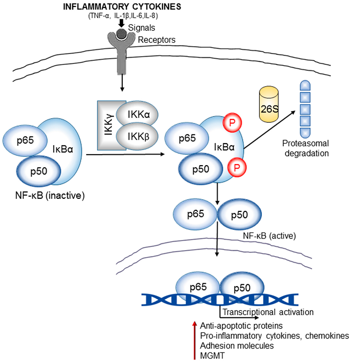
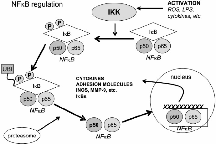
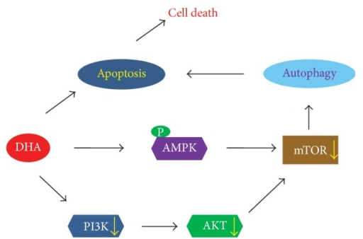
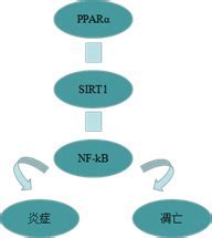
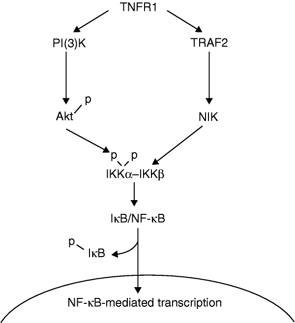
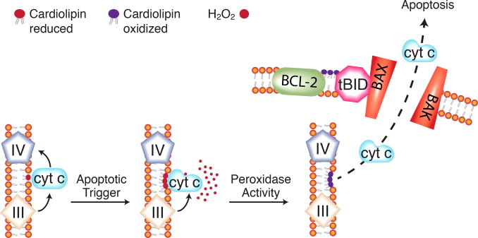

.jpg)
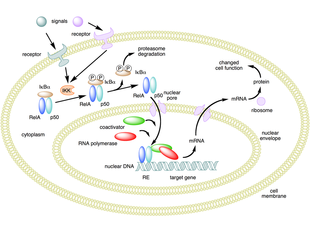
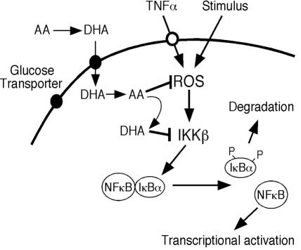
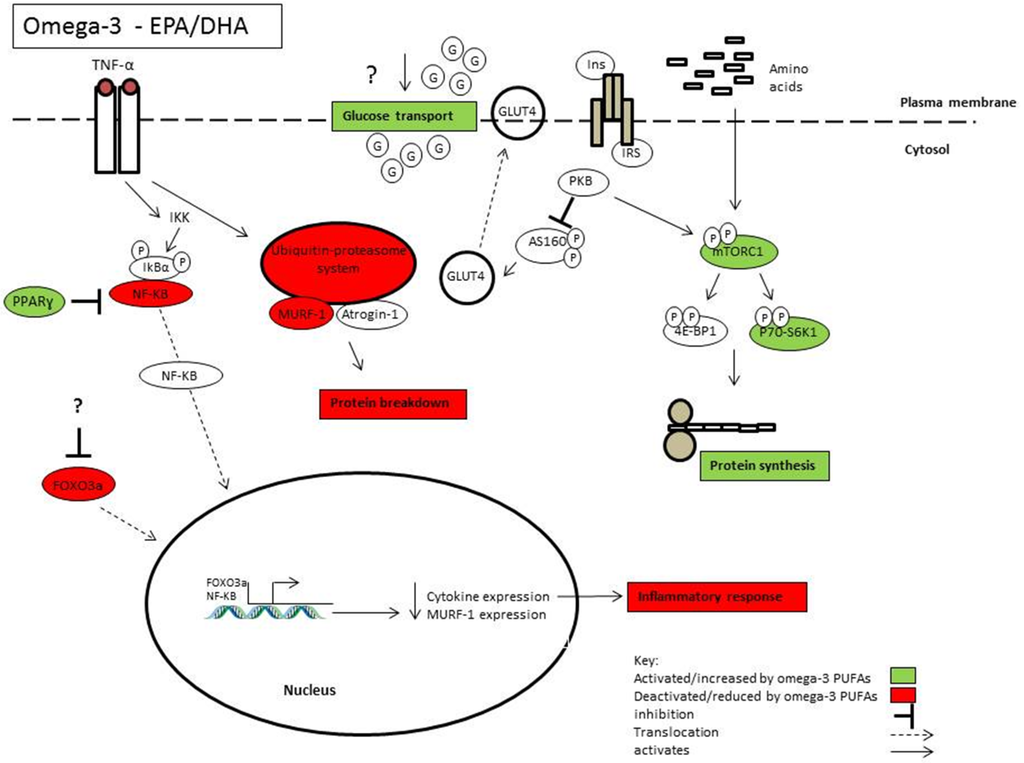
.jpg)

.jpg)
